Download

1 / 54
820 likes | 1.93k Vues
Introduction to Neuroimaging. Dr Mohamed El Safwany , MD. Intended Learning Outcomes. The student should be able to recognize an introduction to neuroimaging. Neuroimaging Modalities. Magnetic Resonance (MR) MR Angiography/Venography (MRA/MRV) Diffusion and Diffusion Tensor MR

E N D
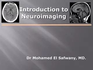
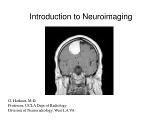
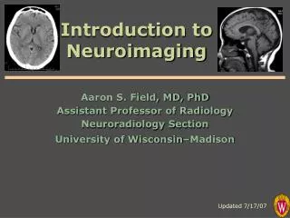
Introduction to Neuroimaging Dr Mohamed El Safwany, MD.
Intended Learning Outcomes • The student should be able to recognize an introduction to neuroimaging.
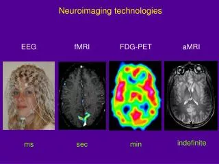
Neuroimaging Modalities • Magnetic Resonance (MR) • MR Angiography/Venography (MRA/MRV) • Diffusion and Diffusion Tensor MR • Perfusion MR • MR Spectroscopy (MRS) • Functional MR (fMRI) • Nuclear Medicine • Positron Emission Tomography (PET) • Radiography (X-Ray) • Fluoroscopy (guided procedures) • Angiography • Diagnostic • Interventional • Myelography • Ultrasound (US) • Gray-Scale • Color Doppler • Computed Tomography (CT) • CT Angiography (CTA) • Perfusion CT • CT Myelography “Duplex”
Radiography (X-Ray) • Disorders of spine: • Trauma • Degenerative Disorders • Post-opeerative
Fluoroscopy (Real-Time X-Ray) • Fluoro-guided procedures: • Angiography • Myelography
Fluoroscopy (Real-Time X-Ray) Digital Subtraction Angiography
Fluoroscopy (Real-Time X-Ray) Digital Subtraction Angiography
Fluoroscopy (Real-Time X-Ray) Myelography Lumbar or cervical puncture Inject contrast intrathecally with fluoroscopic guidance Follow-up with post-myelo CT (CT myelogram)
US transducer carotid Ultrasound
Ultrasound Indications: • Carotid stenosis • Vasospasm - Transcranial Doppler (TCD) • Infant brain imaging (open fontanelle = acoustic window) • Noninvasive, well-tolerated, readily available, low cost • Quantitates blood velocity • Reveals morphology (stability) of atheromatous plaques • Severe stenosis may appear occluded • Limited coverage, difficult through air/bone • Operator dependent Advantages: Disadvantages:
Ultrasound – Gray Scale Gray-scale image of carotid artery
Ultrasound – Gray Scale Plaque in ICA Gray-scale image of carotid artery
Ultrasound - Color Doppler Peak Systolic Velocity (cm/sec)ICA Stenosis (% diameter) 125 – 225 50 – 70 225 – 350 70 – 90 >350 >90
Computed Tomography A CT image is a pixel-by-pixel map ofX-ray beam attenuation(essentiallydensity)inHounsfield Units (HU) HUwater = 0 Bright = “hyper-attenuating” or “hyper-dense”
Computed Tomography Typical HU Values: Air –1000 Fat –100 to –40 Water 0 Other fluids (e.g. CSF) 0–20 White matter 20–35 Gray matter 30–40 Blood clot 55–75 Calcification >150 Bone 1000 Metallic foreign body >1000 Brain
Computed Tomography “Soft Tissue Window” “Bone Window”
Computed Tomography Scan axially… …stack and re-slice in any plane “2D Recons”
CT Indications • Skull and skull base, vertebrae • (trauma, bone lesions) • Ventricles • (hydrocephalus, shunt placement) • Intracranial masses, mass effects • (headache, N/V, visual symptoms, etc.) • Hemorrhage, ischemia • (stroke, mental status change) • Calcification • (lesion characterization)
Skull and skull base, vertebrae Fractures
Ventricles Hydrocephalus
Intracranial masses, mass effects Solid mass Cystic mass
Intracranial masses, mass effects L hemisphere swelling Generalized swelling
Acute Hemorrhage Intraparenchymal Subarachnoid Subdural Epidural
Acute Ischemia Loss of gray-white distinction and swelling in known arterial territory
Calcification Hyperparathyroidism
CT Angiography • Rapid IV contrast bolus • Dynamic scanning during arterial phase • Advanced 2D and 3D Reconstructions: • 2D multi-planar (sagittal, coronal) • Volume–rendered 3D recons
CT Angiography - Head Circle of Willis Vascular Malformations Aneurysms
CT Angiography - Neck Carotid bifurcations Vertebral arteries Aortic arch
CT Angiography - Indications • Atherosclerosis • Thromboembolism • Vascular dissection • Aneurysms • Vascular malformations • Penetrating trauma
CBV CBF MTT CT Perfusion
Hemodynamic Parameters Derived From Concentration-Time Curves Bolus arrival Vein Artery
Hemodynamic Parameter Maps Transit Time (sec) Blood Flow (mL/min/g) Blood Volume (mL/g)
CT Myelography • Spinal CT immediately following conventional myelogram • Cross-sectional view of spinal canal along with spinal cord and nerve roots • Assess spinal stenosis/nerve root compression (e.g. disc herniation, vertebral fracture, neoplasm)
Magnetic Resonance (MR) Hydrogen proton in water or fat MRI
Magnetic Resonance Imaging Transmitter Receiver RF COMPUTER RF = Radio Frequency energy Received signal magnetic field
“T1-weighted” “T2-weighted” w/ fat suppression Magnetic Resonance
Magnetic Resonance T1 T2 Arachnoid Cyst
Diffusion MR Imaging NORMAL CYTOTOXIC EDEMA (Acute Ischemia) Diffusion MR Signal
Magnetic Resonance Imaging Diffusion DWI Highly sensitive to acute ischemia— + within a few hours! No other imaging is more sensitive to acute ischemia although perfusion imaging reveals hypoperfused tissue at risk for ischemia Acute left MCA infarction
Magnetic Resonance Angiography Axial “source” images… …reformatted to “maximum intensity projections” (MIP) Multiple projections allow 3D-like display No need for IV contrast!
Magnetic Resonance Angiography with Perfusion MR MRA Perfusion MR
Magnetic Resonance Tissue contrast in MR may be based on: • Proton density • Water/fat/protein content • Metabolic compounds (MR Spectroscopy) • e.g. Choline, creatine, N-acetylaspartate, lactate • Magnetic properties of specific molecules • e.g.Hemoglobin • Diffusion of water • Perfusion (capillary blood flow) • Bulk flow (large vessels, CSF)
IV Contrast in Neuroimaging • CT: Iodine-based • Iodine is highly attenuating of X-ray beam (bright on CT) • MRI: Gadolinium-based • Gadolinium is a paramagnetic metal that hastens T1 relaxation of nearby water protons (bright on T1-weighted images) • Tissue that gets brighter with IV contrast is said to “enhance” (Brightness, in and of itself, is not enhancement!) • Enhancement reflects the vascularity of tissue, but… • The blood-brain barrierkeeps IV contrast outof the brain! • Enhancement implies BBB is absent or dysfunctional • Remember: Some brain anatomy lives outside the BBB
Enhancement T1 T1+C Hemorrhagic melanoma metastasis