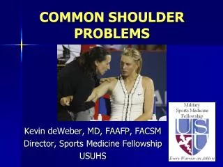
COMMON SHOULDER PROBLEMS
COMMON SHOULDER PROBLEMS. Kevin deWeber , MD, FAAFP, FACSM Director, Sports Medicine Fellowship USUHS. Objectives. Review anatomy Makes for better diagnoses Discuss common shoulder problems Describe current treatments. Anatomy. Scapula Glenoid Acromion Coracoid Subscapular fossa

COMMON SHOULDER PROBLEMS
E N D
Presentation Transcript
COMMON SHOULDER PROBLEMS Kevin deWeber, MD, FAAFP, FACSM Director, Sports Medicine Fellowship USUHS
Objectives • Review anatomy • Makes for better diagnoses • Discuss common shoulder problems • Describe current treatments
Anatomy • Scapula • Glenoid • Acromion • Coracoid • Subscapular fossa • Scapular spine • Supraspinous fossa • Infraspinous fossa
Anatomy • Bursae • Subacromial (Subdeltoid) • Subscapular
Joints of the Shoulder • Acromioclavicular • Glenohumeral • Sternoclavicular • Scapulothoracic • Not a “true” joint
Movement control • Flexion: Pectoralis Major, Deltoid (Anterior), Coracobrachialis • Extension: Deltoid (Posterior), Teres Major • Abduction: Deltoid, Supraspinatus • Adduction: Pectoralis Major, Latissimus, Subscapularis, Infrapspinatus, Teres Minor • Internal Rotation: Subscapularis, Pectoralis Major, Deltoid (A), Latissimus • External Rotation: Infraspinatus, Teres Minor, Deltoid
Inspection Palpation Range of Motion Strength Neuro-Vascular Special Tests Shoulder: Physical Exam
Range of Motion • Forward flexion: 160 - 180° • Extension: 40 - 60° • Abduction: 180◦ • Adduction: 45 ° • External rotation: 80 - 90 ° • Internal rotation: 60 - 90 °
Strength Testing • Rotator Cuff Muscles • S – Supraspinatus • I – Infraspinatus • t - Teres minor • S- Supscapularis • Abduction: Supra • IR: subscap • ER: infra, TM • Other muscles • Deltoid • Biceps • Pecs • Scapular stabilizers
Anatomy • Muscles • Deltoid • Trapezius * • Rhomboids * • Levator scapulae * • Rotator cuff • Teres major • Biceps • Pectoralis muscles * • Serratus anterior * • * Scapular stabilizers
Common Shoulder Problems Instability Impingement Rotator cuff tears AC joint sprains and degeneration Adhesive capsulitis Labral tears Biceps tendinopathy Clavicle fractures
Glenohumeral Instability • DEFINITION: painful feeling of slippage, looseness, “going in and out”
Instability Eval: “FEDS” • Frequency • 1-times • 2-5 • “frequent” >5 • Etiology: Traumatic vs. Atraumatic • Direction (predominant) • anterior • posterior • inferior • Severity: Dislocation vs. Subluxation
Dislocation: impact to externally rotated, abducted arm Acute findings: prominent acromion, anterior fullness Special Tests: Apprehension, Relocation Anterior Instability
Bankart Lesion Anterior capsule torn Anteroinferior labrum torn Recurrent dislocations likely Hill-Sachs Lesion Humeral compression fracture Anterior Dislocation Injuries
Dislocations: Electrocutions, Seizures Acute findings: internal rotation, adduction Special tests: Posterior drawer Load-shift Posterior Instability
Usually atraumatic Special tests: Sulcus sign Inferior Instability
4-view Radiographs: AP Axillary scapular “Y” AC joint MRI Instability Imaging
Attempt ASAP Intra-articularLidocaine HELPS! Use 2-3 techniques until successful Failure: to ER sedation Anterior Dislocation Reduction
Anterior Dislocation Treatment • Referral to Ortho & PhTh • Surgery for younger/athletic patients • Rehabilitation for others • Immobilization • Sling
Definition: compression of the rotator cuff in the subacromial space Symptoms: Pain with Overhead position or flexion/Internal Rotation Anterior, lateral shoulder pain Night Pain Risk Factors: Overhead activities Micotrauma GH Instability Shape of Acromion DJD Impingement
Neer: full Flexion “Neer to the Ear” Hawkins: Internal Rotation Impingement screening tests
Full Can Test: Resistance applied in forward flexion and abduction (SCAPULAR PLANE) Impingement confirmatory test
5cc 1% lidocaine 25-27g needle Postero-laterally Wait 10 minutes for result >50% pain reduction confirms Neer test: Subacromial Injection relieves pain
Impingement • Imaging not initially needed • 4-view shoulder series • MRI if considering surgery • Failed rehab • Pain with ADLs
Acute Phase: Avoid Exacerbating Factors Control Pain/Inflammation Physical Therapy Corticosteroid Injection Recovery Phase: ROM, Strength, Proprioception Maintenance Phase: Longer, Intense Workouts Surgical Intervention: Failed Conservative Measures, Signifcant Disability Impingement Treatment
Similar presentation as Impingement Failed rehab for impingement Persistent pain/weakness after Neer injection test Imaging: x-rays, MRI Rotator Cuff Tears
Rotator Cuff Tear Exam • Supraspinatus: • drop-arm test • Infraspinatus or Teres Minor • External rotation lag sign • Subscapularis • Belly press test
Rotator Cuff Tears • Treatment • Conservative: Similar to Impingement • Surgical: • Young patient, large tears, dominant arm • Failed Conservative Therapy • High-Level Athlete • Unable to perform vocational activities • Success depends upon degree of tendon damage and degeneration
Prolotherapy for RCTs • 25% Dextrose • Platelet-Rich Plasma (PRP) • Concentration of platelets and their growth factors • Process: (30 minutes) • 20-60cc blood is drawn, then centrifuged to produce 3-6ml of PRP • Ultrasound-guided injection
Mechanism: Fall on shoulder Presentation: superior shoulder pain Exam: AC jt TTP +/- deformity or swelling Cross-chest (“scarf”) test AC Joint Sprain
Cross Chest (“scarf”) Test Active Compression (“AC) test AC Joint Sprain
Imaging Bilateral AP Zanca View 10-15 degrees of cephalic tilt Axillary View Evaluates clavicular displacement AC Joint Sprain
AC Joint Sprain: Treatment • Grade I and II: Conservative • Immobilization • Ice, Analgesics • ROM, Strengthening • Anesthetic injection if rapid RTP needed • Grade III: Controversial; refer to Ortho for counseling • Immobilization for up to 4 weeks • Most studies indicate conservative treatment is better • Surgical management with higher rate of complications1 • Conservative management with mean time of 2.1 weeks to return to work2 • Grade IV-VI: Surgical • Taft TN, et al. Dislocation of the acromioclavicular joint. An end-result study. J Bone Joint Surg Am 1987 Sep;69(7):1045-51. • Auwojtys EM; Nelson G. Conservative treatment of Grade III acromioclavicular dislocations. SOClin Orthop Relat Res. 1991 Jul;(268):112-9.
AC Joint Arthritis • Chronic pain at AC joint • Exam: ACJ ttp, + scarf test, + active compression test • X-rays: narrowed AC jt, +/- osteophytes • Tx: • Avoid painful activities • Steroid injections • Surgical removal of distal clavicle (Mumford)
Painful restriction of active and passive GH ROM Risk Factors Idiopathic Diabetes Mellitus Female Gender Ages 40-60 Immobilization Inflammation Stroke Adhesive Capsulitis
Stage I 1-3 months Pain with normal ROM Stage II: “Freezing” 3-9 months Pain and progressive ROM restriction Stage III: “Frozen” 9-15 months Severe ROM restriction with decreased pain Stage IV: “Thawing” 15-24 months Progressive restoration of ROM Adhesive Capsulitis
Adhesive Capsulitis: Treatment • Anti-Inflammatories • ROM, Stretching • Steroid injection into subacromial space or GH jt • Surgical • Dilatation • Manipulation
Causes: Traction Injuries, FOOSH, Overhead motion overuse, MVA Trauma Locations: Superior Labral Anterior-Posterior (SLAP) tear Posterior Anterior (from dislocation) Labral Tears
History: Pain with overhead or cross-body activity Popping, clicking, catching 85% incidence of coexisting pathology Physical (none diagnostic): Crank Test Anterior Slide Test Yegason Test Labral Tears
Type 1: Fraying Injury Type 2: Biceps tendon detached Type 3: “Bucket-handle” tear Type 4: “Bucket-handle” with Biceps detached SLAP Tears
Labral Tears • Diagnostic: Radiograph, MR arthrogram • Treatment: • Physical Therapy for > 3 months • Usually don’t heal. Aim for PAIN CONTROL • Surgery: • Types I and III: Debridement • Types II and IV: Debridement and Reattachment • Post-Op Rehabilitation • Immobilize for 3 weeks • Progress with AROM • Return to full activity after 12-14 weeks
Rarely seen in isolation Labral tears Rotator cuff tears Impingement Exam findings non-specific Biceps Tendinopathy