Download
1 / 35
350 likes | 483 Vues
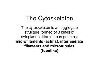
The Eukaryotic Cytoskeleton Oct 9 and 11. Organizing compartments, and a whole lot more!. Typical animal cell. Lodish et al, Fig 5-42. Endoplasmic Reticulum. Lodish et al, Fig 5-47. Golgi’s Apparatus. Lodish et al, Fig 5-49. The Secretory Pathway. Lodish et al, Fig 5-48.

E N D
The EukaryoticCytoskeletonOct 9 and 11 Organizing compartments, and a whole lot more!
Typical animal cell Lodish et al, Fig 5-42
Endoplasmic Reticulum Lodish et al, Fig 5-47
Golgi’s Apparatus Lodish et al, Fig 5-49
The Secretory Pathway Lodish et al, Fig 5-48
Lysosomal Degradation Lodish et al, Fig 5-44
Protein Transport: Overview Lodish et al, 17-50
Budding Fusion Transport Lodish et al, Fig 17-51, 17-59
The eukaryotic cytoskeleton Lodish et al, Fig 19-50
Microtubules Lodish et al Fig 19-1, 19-2
The MTOC Lodish et al Fig 19-5
Microtubule assembly Lodish et al, Fig 19-7
Dynamic Instability Lodish et al Fig 19-14
Melanophore Motility Lodish et al Fig 19-21
Kinesin Lodish et al, Fig 19-23
Dynein Vesicle Transport Lodish et al Fig 19-25
Flagellar Dyneins Lodish et al Fig 19-30, 19-31
Flagellar Beating Lodish et al, Fig 19-27
The Flagellar Machine Lodish et al, Fig 19-29
Flagellar Assembly Lodish et al Fig 19-33
Microfilaments Lodish et al, Fig 18-2
Polarity (S1 decoration) Lodish et al fig 18-3, 18-13
Localization Lodish et al, Fig 18-1
Myosin Lodish et al Fig 18-24
Myosin also moves vesicles Bundle of microfilaments Lodish et al Fig 18-40
Muscle Lodish et al Fig 18-24
The Sliding Filament Hypothesis Lodish et al Fig 18-29
Controlling Contractions Via Troponin/ Tropomyosin (in vivo) Lodish et al Fig 18-32, 18-33a
Intermediate filaments Keratin Lodish et al Fig 19-51
Connecting it all: The Macrotrabecular Lattice Lodish et al Fig 19-54