Download
1 / 60
660 likes | 957 Vues
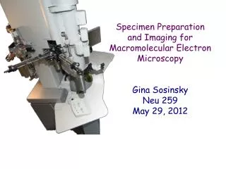
Specimen Preparation and Imaging for Macromolecular Electron Microscopy Gina Sosinsky Neu 259 May 29, 2012. Topics. Range of sample sizes studied by macromolecular microscopy Types of samples (Repeating assemblies) Never-ending quest for higher resolution What limits resolution?

E N D
Specimen Preparationand Imaging forMacromolecular Electron Microscopy Gina Sosinsky Neu 259 May 29, 2012
Topics • Range of sample sizes studied by macromolecular microscopy • Types of samples (Repeating assemblies) • Never-ending quest for higher resolution • What limits resolution? • Techniques • Metal shadowing • Negative/positive staining • Embedding in sugars or tannic acid • Cryo-Electron microscopy • Cryo-Negative staining • Cryo-tomography • Low dose microscopy
Range of Sample Sizes Studiedby Macromolecular Microscopy Paramecium bursariaChlorella Virus 1~1900 Å, ~1 GDa Caulobacter crescentus~0.6 X 2 µm Bacteriorhodopsin~58 Å, ~26 kDa 70S E. coli ribosome~250 Å Theoretical Biophysics Group Beckman Institute University of Illinois at Urbana-Champaign Timothy S. Baker Group UCSD Briegel, et al. (2006) Courtesy J. Frank 100 Å 1000 Å 1 µm
Types of Samples Studiedby Macromolecular Microscopy Repeating Assemblies 70S E. coliribosome Hepatitis B virus core Actin-myosin filament Light-harvesting 2D crystal Single particles with little or no symmetry Single particles with icosahedral or other symmetries Helical symmetry Two-dimensional crystals Baker and Henderson (2001)
Types of Samples Studiedby Macromolecular Microscopy Cells, Organelles, Pleomorphic Viruses, etc. Human Immunodeficiency Virus - 1Zhu, et al. (2006) Baker and Henderson (2001) Nuclear Pore ComplexBeck, et al. (2007)
The Never-Ending Quest for Higher Resolution in EM Points of Resolution in Structural Information of Proteins Fujiyoshi (1998) Adv. Biophys 35:25-80
Limits of Resolution of Various Imaging Technologies The Never-Ending Quest for Higher Resolution in EM & NMR Currently achieved resolution Leis, et al. (2009) Prediction for resolution improvement
The Never-Ending Quest for Higher Resolution in EM Resolution of Selected Solved Structures
The Never-Ending Quest for Higher Resolution in EM Visualizing Helices in Penicillium stoloniferum Virus ~ 350 Å Diameter~7.3 Å resolution X-eyed Stereo
What Limits Resolution? • The vacuum of the microscope - DEHYDRATION! • ~9 X 10⁻⁸ Torr or 1 X 10⁻¹⁰ atm • Look at the lengths we go to avoid this! • We dehydrate it with solvents • We flash freeze it and then freeze dry it • We embed it in heavy metals • We vitrify it (Flash frozen in amorphous ice)
What Limits Resolution? • The vacuum of the microscope - DEHYDRATION! • Lack of contrast - BIOLOGICAL SAMPLES JUST DON’T DO A VERY GOOD JOB AT SCATTERING ELECTRONS!
What Limits Resolution? • The vacuum of the microscope - Dehydration • Lack of contrast - Biological samples just don’t do a very good job at scattering electrons • Radiation damage - Biological samples just don’t like getting hit by the electron beam
Radiation Damage - Primary and Secondary Effects • Primary Effects • Temperature independent • Occurs in the first 10-14 seconds • Excitation of orbital electrons of the sample forms ions and radicals • Secondary Effects • Secondary effects are what cause damage in the sample • Secondary effects are temperature dependent • Chemical and physical changes (breaking C-H and C-C bonds) • Mass loss (can be as great as 50%) • Charging effects • Contamination • Residual hydrocarbons in the vacuum chamber can break into fragments that become deposited on specimens. • Water vapor from insertion of the sample and from photographic film • Loss of order in crystalline specimens
Radiation Damage - Loss of Order in Crystalline Specimens Changes in the electron diffraction pattern of frozen-hydrated catalase crystals resulting from radiation damage <1 e-/Å2 2.5 e-/Å2 2.8 Å 5.0 e-/Å2 11 e-/Å2 Example Dosage: The minimum dosage necessary to see an image on the screen at 20kX magnification is ~4e-/Å2/sec. 8.5 Å Taylor and Glaeser (1976) J. Ultrastruc. Res. 55:448
10 e-/Å2 20 e-/Å2 30 e-/Å2 40 e-/Å2 Radiation DamageFrozen-hydrated Simian Virus 40 Dose Series
Techniques • Metal shadowing • Negative/positive staining • Embedding in sugars and tannic acid • Cryo-electron microscopy • Cryo-negative staining • Cryo-tomography
Techniques - Metal Shadowing Wischnitzer (1970) Introduction to electron microscopy, 2nd ed.
Techniques - Metal Shadowing • Problems • Only see the surface • Standard techniques are low resolution, evaporated metals tend to be granular • Metal decoration Actin + S1, Courtesy of John Heuser
Techniques - Negative/Positive Staining • “Negative stain” is a misnomer. Most of the stain fills in sample depressions thereby preventing sample collapse. It does not stain the sample. • Sample appears “white” and the electron-dense stain is “black”. • Helps to reduce dehydration and radiation damage effects. • Attainable resolution is ~ 15-25 Å (~10 Å - GroEL De Carlo et al. 2008). 8 Å for crystals. • Positive staining occurs when ions of the stain react with the molecule. Hayat & Miller (1990)Negative Staining
Techniques - Negative/Positive Staining Examples of Commonly Used Negative Stains • Others: • Methylamine tungstate • Silver nitrate • Aurothioglucose • Sodium tetraborate • Cadmium iodide
Techniques - Negative/Positive Staining Choosing the Proper Stain • High density • 3.8 - 5.7 gm/cc versus 1.37 gm/cc for protein • High solubility • High melting and boiling points • Stability in the beam • Stain needs to have a fine granularity (0.4 to 1.5 nm) • No chemical reaction with the specimen • Choice of stain may depend on the pH and salt concentration needs of the sample
Carbon-filmed grid Techniques - Negative/Positive Staining Procedure - Setup dH2O Sample Filter paperwedges 1% UA stain in water Grid Box
Techniques - Negative/Positive Staining Procedure - Hydrophilic Carbon Surface Hydrophobic surface Hydrophilic surface
Techniques - Negative/Positive Staining Procedure - Glow Discharging the Grids
Techniques - Negative/Positive Staining Procedure - Apply Sample to Grid
Techniques - Negative/Positive Staining Procedure - Wash with dH2O
Techniques - Negative/Positive Staining Procedure - Apply Stain
Techniques - Negative/Positive Staining Procedure - Blot with Filter Paper
Techniques - Negative/Positive Staining Results Bacteriophage T4 Maize Streak Virus
Techniques - Negative/Positive Staining Problems • Unpredictable and uneven staining • High contrast images only the surface • Sample flattening • The electron beam can redistribute the stain • Different stains give different views Parmecium bursariaChlorella virus Uranyl acetate stained Cryo-electron microscopy
Techniques - Negative/Positive Staining Artifacts “The question of what is artifact and what is not is a persistent one in electron microscopy, especially when micrographs depict what are essentially newer unexplored structures.“ Dr. Keith Porter, 1979. From Heuser (2002)
Techniques - Embedding inSugars or Tannic acid • Making the preparation is similar to that of negative staining. • Specimen is supported by a matrix of concentrated sugar to maintain the need for hydration. • Often used with crystalline samples in order to keep them flat on the grid • Very beam sensitive, often need to keep sample at liquid nitrogen temperature • Very low contrast, sugar scatters electrons as well as protein (contrast matching)
Techniques - Cryo-Electron Microscopy (CryoEM) • Specimens are frozen in non-crystalline (vitreous ice). Freezing must be done in less than 10-4 sec. • Specimens are “frozen-hydrated”. This overcomes the problem of putting a hydrated sample in a vacuum. • Specimens are observed with “native contrast” - no staining, no fixatives. • Samples must be maintained below ~-140o C in the microscope. (below the vitreous to crystalline ice phase transition) • Maintains the native structure of the molecule to atomic resolution. Bacteriophage 29
Techniques - CryoEM Holey Carbon Support Film
Techniques - CryoEM Sample Preparation Equipment
Techniques - CryoEM Sample Preparation Equipment Guillotine Plunger EM tweezers LN2 dewar Foot switch
Techniques - CryoEM Sample Preparation Equipment Guillotine Plunger ethane EM forceps EM grid LN2 LN2 dewar
Techniques - CryoEM Addition of Sample and Blotting
Techniques - CryoEM Addition of Sample and Blotting Filter paper Filter paper EM grid Sample
LN2 (-196 °C) EM grid ethane Techniques - CryoEM Plunging the Grid into Ethane and Transfer to Nitrogen
Techniques - CryoEM Transferring to the Grid Storage Box
Techniques - CryoEM Automated Freezing with the FEI Vitrobot Environmental chamber Computer-controlled
Techniques - CryoEM Using the Vitrobot
EM tweezers Sampleon grid Filter paper disks Techniques - CryoEM Using the Vitrobot
Techniques - CryoEM Using the Vitrobot
Techniques - CryoEM Using the Vitrobot
Techniques - CryoEM Cryo-transfer Workstation and Holder Cover Transfer Area Cryo-Shield Nitrogen Dewar Workstation
Techniques - CryoEM Transferring the Grid into the Cryo-holder Grid Box Cryo-Shield Grid Clipring
Techniques - CryoEM Transferring the Grid into the Cryo-holder Grid Box Cryo-Shield Grid Clipring
Techniques - CryoEM Transfer of the Grid to the Cryo-holder