Download
1 / 24
250 likes | 428 Vues
The Biology of Keratins or why are they so important?. Birgit Lane birgit@cmm.a-star.edu.sg CRUK Cell Structure Research Group, University of Dundee and Centre for Molecular Medicine, Singapore. “ We live our whole lives less than 1/10mm away from certain death “. W. Montagna.

E N D
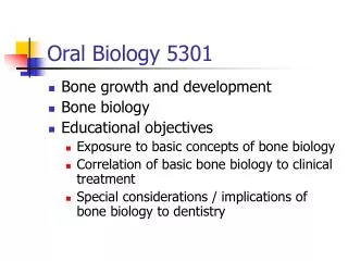
The Biology of Keratinsorwhy are they so important? Birgit Lane birgit@cmm.a-star.edu.sg CRUK Cell Structure Research Group, University of Dundee and Centre for Molecular Medicine, Singapore
“ We live our whole lives less than 1/10mm away from certain death “ W. Montagna immunofluorescence
Skin - the ultimate barrier tissue • Tough • Waterproof • Disposable • Self-healing • Site of most cancers EPIDERMIS K1/K10 K5/K14 DERMIS
Scratch wound assays movie: Sharon Jenner
Keratins as Intermediate filament proteins Cytoskeleton filaments Many family members Major structural components of sheet tissues Different proteins in different body parts Many diseases (40+)
The Intermediate Filament Gene Family Type I keratins epithelia Type II keratins epithelia 54 Type III vimentin-like mesenchyme Type IV neurofilaments neurones Type V lamins all nuclei Type VI (variable) ______________________________ Total 70 }
Structure of intermediate filaments after Herrmann and Aebi
monomer dimer tetramer unit length filament immature filament 10 nm filament
Type II Type I K2p K9 K1b K9- thick dry: palmoplantar skin K1 K10 K2e Thin dry: cornified (skin) K3 K12 Transparent: cornea stratified (barrier) K13 K4 Wet: mucosal K6a K16 Stress: fast turnover, wound response K6b K17 K15 Basal cells K5 K14 basal K19 Simple: internal, secretory, embryonic K8 K18 simple K20 K7 Keratin filament proteins
EPIDERMIS K1/K10 K5/K14 DERMIS
Type II Type I K2p K9 K1b K9- thick dry: palmoplantar skin K1 K10 K2e Thin dry: cornified (skin) K3 K12 Transparent: cornea stratified (barrier) K13 K4 Wet: mucosal K6a K16 Stress: fast turnover, wound response K6b K17 K15 Basal cells K5 K14 basal K19 Simple: internal, secretory, embryonic K8 K18 simple K20 K7 Keratin filament proteins
Bullous congenital ichthyosiform erythroderma (BCIE, EHK) • Thickened skin • Fragile suprabasal cells • Mutations in K1 or K10 • Mostly rod end hotspots
Type II Type I K2p K9 K1b K9- thick dry: palmoplantar skin K1 K10 K2e Thin dry: cornified (skin) K3 K12 Transparent: cornea stratified (barrier) K13 K4 Wet: mucosal K6a K16 Stress: fast turnover, wound response K6b K17 K15 Basal cells K5 K14 basal K19 Simple: internal, secretory, embryonic K8 K18 simple K20 K7 Keratin filament proteins
Ichthyosis bullosa of Siemens (IBS) • Fragility late in differentiation • K2e, K1 mutations • Mostly rod end hotspots
Effects of keratin mutations on cell integrity None K5/K14 K1/K10 K2e EBS BCIE (EHK) IBS
Type II Type I K2p K9 K1b K9- thick dry: palmoplantar skin K1 K10 K2e Thin dry: cornified (skin) K3 K12 Transparent: cornea stratified (barrier) K13 K4 Wet: mucosal K6a K16 Stress: fast turnover, wound response K6b K17 K15 Basal cells K5 K14 basal K19 Simple: internal, secretory, embryonic K8 K18 simple K20 K7 Keratin filament proteins
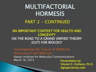
Pachyonychia congenita - Type 1 (K6a, K16) - Type 2 (K6b, K17)
K6a, K16 - PC-1 K6b, K17 - PC-2 major keratins in “wet” skin surfaces; outer hair follicle sheath; nail bed. NOT normally in thin “dry”skin but more in thick skin INDUCED by trauma, wounding Often expressed in tissue cultured cells Small amounts in “wet” epithelia. Lots in deep hair follicle sheath; some basal cells; nail bed. Specialized and unspecialized cells. NOT normally in thin “dry” skin. INDUCED by trauma, wounding, inflammation Often expressed in tissue cultured cells K6a/K16, K6b/K17 - the stress response keratins
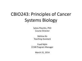
+ - K16 induced by epidermal wounding K16 Healing blister: Keratinocytes downregulate K10 and express K6/K16/K17 upon stress or injury.
epidermis Outer hair follicle: lots of K16, very little K17 Deep hair follicle: lots of K17, very little K16
Disorders caused by defective intermediate filaments (at least 40 disorders so far) http://www.interfil.org