Download
1 / 1
10 likes | 176 Vues
Green Fluorescent Protein. DEVD. M2. 36. 27. 20. Yellow Fluorescent Protein. DEVD. M2. Cerulean Fluorescent Protein. Cyan Fluorescent Protein. DEVD. DEVD. M2. M2. 37. 25. 20. 15. 37. 25. 20. - - +. - + -. + - -. + - +. + + -. 15. CA-GFP WT C7 C186A C7. FasL

E N D
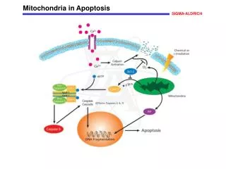
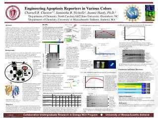
Green Fluorescent Protein DEVD M2 36 27 20 Yellow Fluorescent Protein DEVD M2 Cerulean Fluorescent Protein Cyan Fluorescent Protein DEVD DEVD M2 M2 37 25 20 15 37 25 20 - - + - + - + - - + - + + + - 15 CA-GFP WT C7 C186A C7 FasL TNF- Intrinsic Extrinsic Mitochondrial Stress Procaspase-8 DISC complex Procaspase-3 Procaspase-6 Procaspase-7 Mitochondria Cytochrome c Active Caspase-8 Apaf-1 Apaf-1 + dATP Active Caspase-3 Active Caspase-6 Procaspase-9 Apoptosome complex Active Caspase-7 Apoptosis laminA PARP IAPs ?? Cleavage of Caspase-specific targets SDS-PAGE Full Length Caspase-7 CFP CACFP CFP Anti-Caspase-7 ∆Pro- Caspase-7 Large Subunit (Caspase-7) Uncleaved CA-GFP Anti-GFP Cleaved CA-GFP Engineering Apoptosis Reporters in Various Colors Charnell R. Chasten1,2, Samantha B. Nicholls2, Jeanne Hardy, Ph.D.2 1Department of Chemistry,North Carolina A&T State University, Greensboro, NC 2Department of Chemistry,University of Massachusetts Amherst, Amherst, MA Green Fluorescent Protein DEVD QP Results: Abstract: Apoptosis is a biological process, which is involved in over 50% of all diseases lacking suitable treatment. Apoptosis is dependant on the action of caspase proteases. We have developed ed a fluorescent reporter of apoptosis called Caspase Activatiable-Green Fluorescent Protein (CA-GFP). Prior to cleavage by caspases, CA-GFP is in a “dark” state and has no fluorescence intensity. After caspase cleavage, CA-GFP shifts to a “bright” state. For various applications such as experiments in an array of organisms or in the presence of other fluorescent reporters, additional colors of apoptosis reporters would be useful. Our goal here is to expand the spectrum of apoptosis reporters to include cyan fluorescent protein (CFP), Cerulean fluorescent protein (CerFP) and yellow fluorescent protein (YFP). Using quikchange methods for site-directed mutagenesis we produced a number of vectors to express CA-CFP and CA-YFP. These vectors allow experiments to be conducted both in vitro and in cells to monitor the utility of these new reporters. Both CA-CFP and CA-YFP are in a “dark” state before cleavage and have potential to become fluorescent after cleavage. Inactive caspase CA-YFP is dark prior to caspase cleavage and can be cleaved by caspase-7 CA-CFP is dark prior to caspase cleavage CA-GFP is Dark before cleavage and bright after proteolytic cleavage. Green Fluorescent protein (GFP) is represented in gray in the dark state and green when bright, the caspase-7 cleavage site (DEVD) is in blue and the peptide, which quenches fluorescence is represented in gray. After the Quenching Peptide has been cleaved off by the active caspase the GFP goes from ‘dark’ to ‘bright’ and has a significant increase in fluorescence signal. CFP expressed from the vector pET21b has the expected fluorescent spectrum. The observed excitation and emission wavelengths for the purified protein are 446 nm and 509 nm respectively. Active caspase The absorbance spectrum above shows that before cleavage CA-YFP is dark and has a relatively flat spectrum in comparison to that of YFP which has an absorbance maximum at 516 nm. This indicates an immature chromophore in the CA-YFP. When the QP (quenching peptide) is appended to CFP making CA-CFP in the vector pET21b, fluorescence is silenced. CA-GFP to responds to active caspase-7 protease. CA-GFP is cleaved upon co-expression with caspase-7 in E.coli. As a control, CA-GFP was also co-transformed with an inactive mutant of caspase-7 (C186A) in which the active site. Only when CA-GFP was co-expressed with active caspase-7 was robust GFP fluorescence observed. The increase in fluorescence is on average 50-fold greater with wild-type caspase-7 than with the inactive mutant. CA-YFP alone 0 5 10 20 30 60 120 O/N Background: Apoptosis is the mechanism by which cells commit suicide. Cells are targeted to die under certain disease conditions and as a normal part of organismal development. A tool that can report on apoptosis is valuable both in developmental biology and in assessing potential side effects of candidate drugs that lead to cell death in various tissues or organs. Uncleaved CA-YFP 36 Cleaved CA-YFP 27 Large Subunit (Caspase-7) 20 The Apoptotic Pathways. The two pathways lead to activation of the executioner caspases-3, -6, and -7 resulting in cell death. The executioner caspases recognize the four amino acid sequence DEVD. The quenching of fluorescence in CA-CFP compared to CFP can be observed under visible light (black background) or under fluorescent light (blue background). Small Subunit (Caspase-7) The SDS-PAGE gel above shows the digest of CA-YFP with active Caspase-7. The presence of active caspase-7 leads to cleavage of CA-YFP. Conclusions and Future Directions: We have observed that the CA-FP constructs are in a “dark” state prior to caspase cleavage. These constructs have been changed to a “bright” state by Caspase-7 activation. The CA-CerFP construct’s “dark” state indicates that even faster folding FP variants and those with higher quantum yields can be successfully used in our system. We intend to expand the color pallet of our CA-FP’s to cover a broader portion of the fluorescence and possibly infrared spectra. Further research is going to be done to determine the effectiveness of these reporters in E. coli and mammalian systems. CA-CerFP is dark prior to caspase cleavage and can be cleaved by caspase-7 Changing the color of protease reporter proteins. The three fluorescent proteins that have been chosen as new protease reporters are cyan fluorescent protein (CFP), Cerulean fluorescent protein (CerFP) and yellow fluorescent protein (YFP). To enable co-expression with various proteases we have designed constructs in two different DNA vectors, each with different origins of replication. Different origins of replication and selectable markers allow co-expression in bacterial cells. One vector is pET21b, which contains a colE1 origin of replication and the other is pACYC which has a p15A origin of replication. An expression construct for CA-CFP-pET21b Cerulean fluorescent protein (CerFP) expressed from the vector pET21b has the expected fluorescent spectrum. The observed excitation and emission wavelengths for the purified protein are 439 nm and 484 nm respectively. CA-CerFP was dark similarly to CA-CFP and can be activated by cleavage with Caspase-7. The family of proteases known as caspases are critically important in regulating the process of apoptosis. Due to the involvement of apoptosis in many diseases, caspases are popular drug targets. They are also important in understanding developmental processes as apoptosis is essential for the development of multi-cellular organisms. There are currently several existing caspase activity reporting systems however, they have various disadvantages such as having high background signal, or reporting on a ‘bright to dark’ mechanism. Quikchange site directed and multi site directed mutagenesis enables DNA mutations CA-CerFP alone 0 5 10 20 30 60 120 O/N • Acknowledgments: • The Collaborative Undergraduate Research in Chemistry (CURE) REU program funded by the National Science Foundation. • SN has been funded by the NSF IGERT in Cellular Engineering DGE-0654128 Uncleaved CA-CerFP 36 The heterodimer of caspase-7. The apopototic protease caspase-7 consists of two large (lighter) and small (darker) subunits. Cleaved CA-CerFP 27 20 References: Tsien, Roger. Molecular biology and mutation of green fluorescent protein. Green Fluorescent proteins: properties, application and protocols. 2006. 2, 8-12. Large Subunit (Caspase-7) Objective: The objective of this work is to introduce mutations into the Enhanced Green Fluorescent Protein (EGFP) in the CA-GFP constructs that will change the color of the fluorescence. In this way we will expand the spectrum of colors that can be used as fluorescent protease reporters so that multi-protease imaging can be accomplished within the same cell or organism. Small Subunit (Caspase-7) The amino acids which were mutated to obtain the cyan and yellow variants are shown above along with the mutations. Blue amino acid substitutions were introduced using Quikchange mutagenesis by CRC to make the cyan mutants. Black mutations were introduced by SBN for cerulean and yellow variants. The SDS-PAGE gel above shows the digest of CA-CerFP with active Caspase-7. The presence of active caspase-7 leads to cleavage of CA-CerFP. Collaborative Undergraduate Research in Energy REU Program ● University of Massachusetts Amherst