Correlation of SHH and ERα mRNA Expression in Diffuse and Intestinal-type Gastric Cancer
20 likes | 123 Vues
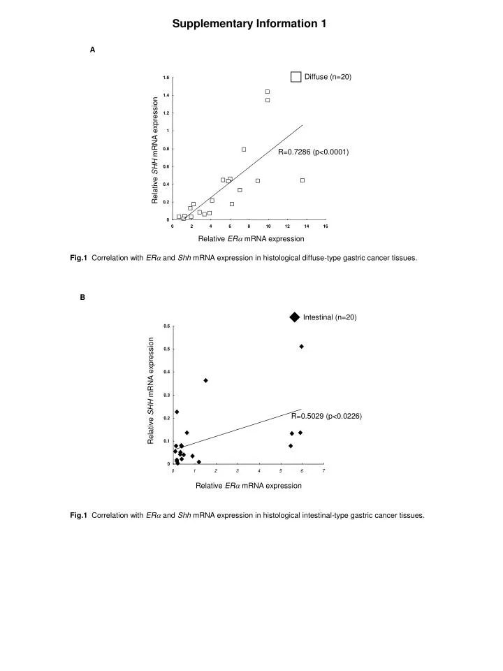
This study investigates the correlation between SHH and ERα mRNA expression in histological tissue samples of gastric cancer. In diffuse-type gastric cancer, a strong positive correlation (R=0.7286, p

Correlation of SHH and ERα mRNA Expression in Diffuse and Intestinal-type Gastric Cancer
E N D
Presentation Transcript
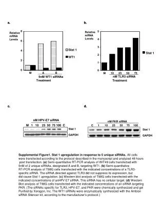
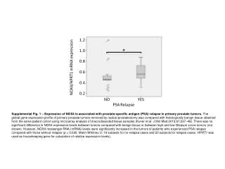
Supplementary Information 1 A Diffuse (n=20) R=0.7286 (p<0.0001) Relative SHH mRNA expression Relative ERa mRNA expression Fig.1Correlation with ERa and Shh mRNA expression in histological diffuse-type gastric cancer tissues. B Intestinal (n=20) Relative SHH mRNA expression R=0.5029 (p<0.0226) Relative ERa mRNA expression Fig.1Correlation with ERa and Shh mRNA expression in histological intestinal-type gastric cancer tissues.
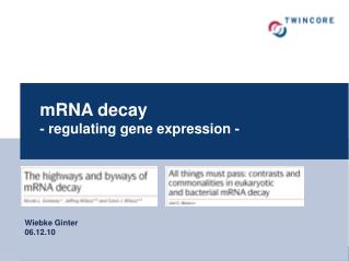
Supplementary Information 2 A NCI-N87 KATO III * Cell number (% of control) Cell number (% of control) E2(nM) E2(nM) TAM (mM) TAM (mM) Fig. 2A Cell proliferation in the presence of E2 alone or in combination with Tamoxifen (TAM) in KATOIII and NCI-N87. 1mM of TAM did not affect the proliferation of ERa-positive gastric cancer cells. Results are expressed as mean ± s.d. *p < 0.05; B MK-1 * Cell number (% of control) E2(nM) TAM (mM) Fig. 2B Cell proliferation in the presence of E2 alone or in combination with Tamoxifen (TAM) in MK-1 cells as ERa-negative cell. 1 mM of TAM did not inhibit the proliferation of MK-1 cells. However, 3mM of TAM inhibited the proliferation of MK-1 cells. Data indicate that 3 mM of TAM may be cytotoxic. Results are expressed as mean ± s.d. *p < 0.05;