Download
1 / 30
300 likes | 549 Vues
Non-membran ous cell organelles. Cytoskeleton – structure, function and tissue specificity. Specialization of the cell surface. Intercellular junctions. Biologic a l motors – m olecular principle s. Institute of Histology and Embryology. Author: Prof. MUDr. Jindřich Martínek, DrSc.

E N D
Non-membranouscell organelles. Cytoskeleton – structure, function and tissue specificity. Specialization of the cell surface. Intercellular junctions. Biological motors – molecular principles Institute of Histology and Embryology Author: Prof. MUDr. Jindřich Martínek, DrSc. Subject: General Histology and general embryology Code: 82241 Date: 2013, October, 10th and 12th
NON-MEMBRANOUS CELL ORGANELLS • NUCLEOLUS • RIBOSOME • CENTRIOLE • CYTOSKELETON thin filaments - actin intermediate filament lamins (A, B, C) cytokeratins (1 – 20) desmin, vimentin nestin microtubules Becker: The world of the cell, 1986
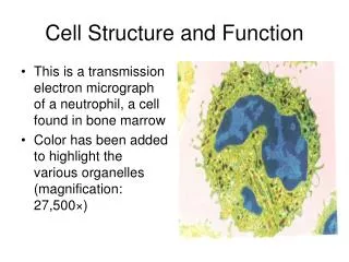
NON-MEMBRANOUS CELL ORGANELLES • NUCLEOLUS– localization in the karyoplasm – nucleolar organizing centers (NORs) - two components (amorphous and granulous) – site of transcription of r-RNAs(ribosomal) – compact, network and ring-form type • CENTRIOLE – specific organization of microtubeles (9 triplets) – MTOC – microtubular organizing center function, simple and multiple centriolar replication. Role in formation of mitotic and meiotic spindle and basal bodies of cilia and flagella • CYTOSKELETON – complex of thin (5-7 nm) and intermediate (10-15 nm) filamentsand microtubules (24 nm) – actin (thin) filaments with myosin and tubulins (of microtubules) with dynein and kinesin represent molecular motors for intracellular traffic – intermediate filaments are nonpolar – nestin, cytokeratins, vimentin, desmin – tissue specific – cells of epithelial origin (cytokeratin), mesenchymal origin (desmin), muscle tissue (vimentin) • RIBOSOMES – (15-30 nm) composed of two subunits (small and large) – play a role in translation – protein synthesis – as free polysomes and membrane bounded (GER)
NUCLELUS – non-membranous cell organellederived from activated parts of some chromosomes – nucleolar organizing regions (NORs) or nucleolar organizing centers (NOCs) Types: compact – initial transcription of rRNA at the single NORs; network – massive transcription in gradually confluent NORs ring-form – decreasing transcription at the single NOR FC = fibrilar center – NOC PF = pars fibrosa – accumulated transcript PG = pars granulosa – ribosomal assembly PNC = perinucleolar chromatin PNC PF PG FC PNC
VISUALISATION OF NUCLEOLAR ORGANISERS USING IMMUNOHISTOCHEMICAL PROBES
TYPES OF NUCLEOLI:a) compact – currently activated nucleolar organiserb) with nucleolonema –massive formation of ribonucleoproteins around NORs - typical network appearancec) ring form –decreased of RNA transcription in the inactivated nucleolar organiser 1 3 2 4 2 3 1 5 6 1 = perinucleolar chromatin 2 = nucleolar organiser 3 = nucleolonemata 4 = nuclear channel system 5 = lamelae annulatae 6 = lysosomes
NUCLEUS RIBOSOMES (15 – 30 nm) free polysomes GER PROTEIN SYNTHESIS NUCLEAR PORES GER GER
RIBOSOME – TRANSLATION MECHANISM – POLYSOMES Start codon (AUG) Currently translating codon (arbitrary)
Scheme of the protein synthesis on free polysomes (Junqueira´s Basic Histology, Mesher, 2010) ribosome GER GER GER mRNA free protein in the cytoplasm Electron micrograph: Histology, Ross, Pawlina 2010 Arrows: free polysomes Rough (Granular) Endoplasmic Reticulum rER (GER)
IMMUNOHISTOCHEMICAL DETECTION OF SOME CYTOSKELATAL COMPONENTS Microtubules (red), actin filaments (green)–fluorescence microscope
CHARACTERISTICS OF CYTOSKELETAL COMPONENETS Ross, Pawlina: Histology, 2006 Actin filaments Intermediate filaments Microtubules Diameter: 6 – 8 nm 10 – 12 nm 24 nm Composition: Polymer of G-actin Various proteins Dimers of α- and ß-tubulin Structure: Double-stranded F-actin helix Ropelike fiber Hollow non-branched cylinder Thin flexible filament Strong, stable structure Exhibit dynamic instability Readily dissociate and reassamble Enzyme activity: ATP hydrolitic activity None GTP hydrolytic activity ATP-dependent polymeration GTP-dependent polymeration Location and function in the cell: Terminal web Extend across cytoplasm Core of cilia and flagellum Zonula adherens connecting desmosomes Centriole Core of microvilli and hemidesmosomes Mitotic spindle Contractile ring in the Nuclear lamina of nucleus Provide network “railroad dividing cell Support of cell processes tracks“ for movement of Contractile elements Provide mechanical strenght organelles within cell of muscles and resistence to shearing Movement cilia and forces chromosomes (during cell division
ACTINthin filaments (5 – 7 nm)G and F actin – ATP dependent (poly- and depolamerization)MOLECULAR MOTOR COMPONENTtogether with myosin Smooth muscle cells Hearth muscle cells - cardiomyocytes
FUNCTION OF MICROFILAMENTS AND MICROTUBULES IN REGULATIONS AND INTRACELLULAR TRAFIC
THE MOLECULAR MOTOR PROTEINS WORKING WITH MICROFILAMENTS (ACTIN) Unipolarly organized myosin (myosin monomers – myosin I) – one way movement of cargo Bipolar organization of myosin (myosin monomers – myosin II) – ontractile activity with opposite directionmovement of actin filaments – muscle tissue Unipolarly working myosin (I) – attached to the cytoplasmic membrane – formation of pseudo- podia movement of cell
THE MOLECULAR MOTOR PROTEINS WORKING WITH TUBULINS OF MICROTUBULES Kinesins move along the MT tothe plus end and can transport cargo (organelles) fromthe cytocentrum toward the cell periphery Dyneins move along the MT to the minus end, transport cargo (endocytotic vesicles) from the cell periphery toward the MTOC. Scheme: Histology, Ross, Pawlina, 2010
INTERMEDIATE FILAMENTS –non-polarlamins A, B, C – nuclear laminacytokeratins – cells of germ layer origindesmin – cells of mesenchymal originvimentin –cells of mesenchymal originneurofilaments – nerve cellsGFAP – glial fibrillary associated protein (acidic) nestin – during cell developmentCYTOKERATIN 18 – fission products – APOPTOTIC MARKER
MICROTUBULES(25 nm) tubulin α, β, γ(GTP – dependent polymerization)MOLECULAR MOTOR COMPONENT – together with dynein (+ -) and kinesin (- +)
CENTRIOLAR PAIR - CENTROSOME CILIUM – KINOCILIUM Ross, Pawlina: Histology, 2006
Centriolar pair = diplosome – centrosome • Centriole: Procentriole: • = 200 nm = 200 nm L = 400 nm L = 200 – 400 nm Centriolar DNA – MTOC – centriolar precursors CENTRIOLES AND CENTRIOLAR REPLICATION MULTIPLE REPLICATION – starts as synthesis of precursor material (tubulins) and continues as induced assembly of nine procentriolar triplets of microtubules – typical for cells with kinociliary apparatus at the apical surface SIMPLE REPLICATION – begins from existing centriolar pair
ZONULA OCCLUDENS – tight junctions – missing intercelluar space Mucus layer ZONULA OCCLUDENS – freeze fracturing technique Ridges Grooves Ciliary necklaces Ross, Pawlina: Histology, 2006
Golgi complex Zonula occludens Zonula adherens Desmosome – macula adherens Cytokeratin – intermediate filaments
ZONULA ADHERENS – terminal web – transverally arranged actin filaments inserting via α-actinin and vinculin and catenin into E-cadherin moleculs
GAP JUNCTIONS – NEXUSSES –communicative cell to cell interconnectionsconnexon – canal formationconnexins – membrane proteins
MICROVILLI • = 0.1 m lenght = 1 – 5 m – core – actin filaments Regularly arranged and numerousmicrovilli represent specific surface specialization of an absorptive epithelium – brush border – at the apical pole of cells. Stereocilium – long and branched microvillus Ross, Pawlina: Histology, 2006
Microvilli at the cell suface and intercellular interconnection ZO = zonula occludens; ZA = zonula adherens; D = desmosome zo ZA D
SOLITARY (single or individual) CILIUM = 0.25 m L = 3 – 5 (7) m (50 for tail of spermatozoon) Consist of basal body (9 triplets – MTOC)and axonemal complex (9 doublets and 1 central pair of microtubules). Basal body develops from one of centrilar pair and therefore it is lo- cated often in the CYTOCENTRUM. Pinocytotic vesicle Ciliary sheat Basal body Axonemal complex Ross, Pawlina: Histology, 2006
KINOCILIARY APPARATUS Cilia and microvilli at the cross section Oviduct – simple columnar ciliated epithelium
CILIA, MICROVILLI, JUNCTIONAL COMPLEXES AND TERMINAL WEB IMMUNOFLUORESCENT DETECTION OF PANCADHERIN IN SEMINIFEROUS TUBULES OF TESTIS (Zonulae adherentes) BETWEEN SERTOLI CELLS PROCESSES INTERDIGITATIONS
BASOLATERAL LABYRINT Typical for absorptive type of epithelial cells as in the proximal tubules (kidney) or in lining of so called striated ductsin some salivary glands. Specific for an intesive Ion transport (energy needed) by e.g. Na+,K+ ATP-ase as an integral protein of the plasma membrane Ross, Pawlina: Histology, 2006 Stevens, Lowe: Histology, 1993 Stevens, Lowe: Histology, 1993