Cell Structure and function
Cell Structure and function. Chapter 4 (in the Rizzo Class Sequence) Chapter 7 in Prentice Hall. Section Outline. Section 7-1. 7–1 Life Is Cellular A. The Discovery of the Cell 1. Early Microscopes 2. The Cell Theory B. Exploring the Cell C. Prokaryotes and Eukaryotes

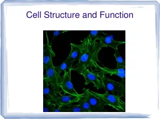
Cell Structure and function
E N D
Presentation Transcript
Cell Structure and function Chapter 4 (in the Rizzo Class Sequence) Chapter 7 in Prentice Hall
Section Outline Section 7-1 • 7–1 Life Is Cellular A. The Discovery of the Cell 1. Early Microscopes 2. The Cell Theory B. Exploring the Cell C. Prokaryotes and Eukaryotes 1. Prokaryotes 2. Eukaryotes
Watch Out!!!! • Keep a watchful eye out for website links some can be used to access labs, animations or videos hosted online.
Section Outline Anton van Leeuwenhoek, 1665 - 75 Anton van Leeuwenhoek, • the person incorrectly given credit for the invention of the microscope • studies organisms living in pond water . • He calls them "Animalcules."
Section Outline Robert Hooke • 1665 - looks at cork under a microscope. • Calls the chambers he see "cells"
Section Outline Schleiden and Schawann 1830 - German scientists summarize the findings of many scientists and conclude that all living organisms are made of cells .
Section Outline Rudolf Virchow 1855- Stated, ”All Cells come from Pre-existing Cells”
Section Outline Cell Theory “All organisms are composed of cells” • The cell is the structural unit of life - units smaller than cells are not alive • The cell is the Functional unit of life • Cells arise by division of preexisting cells - spontaneous generation does not exist
Section Outline Cell Theory 3 facts 3 Exceptions
Bacterial Forms Binary Fission
Prokaryotic vs Eukaryotic Cell membrane Cytoplasm Cell membrane Cytoplasm Prokaryotic and Eukaryotic Cells Prokaryotic Cell Nucleus Eukaryotic Cell Organelles
Prokaryotes vs Eukaryotes Cell membrane Cytoplasm Cell membrane Cytoplasm Prokaryotic and Eukaryotic Cells Section 7-1 Prokaryotic Cell Nucleus Eukaryotic Cell Organelles
Venn Diagrams Prokaryotes Eukaryotes *Complex **Forms tissues ***Has Membrane bound Organelles EX. Nucleus Endoplasmic reticulum Golgi apparatus Lysosomes Vacuoles Mitochondria Cytoskeleton *simpler **Normally single celled Bacteria ***NO (lacks) membrane organelles Cell membrane Contain DNA Cytoplasm Ribosomes
Interest Grabber Division of Labor Section 7-2 • A cell is made up of many parts with different functions that work together. Similarly, the parts of a computer work together to carry out different functions. • Working with a partner, answer the following questions. • 1. What are some of the different parts of a computer? • What are the functions of these computer parts? • 2. How do the functions of these computer parts correspond to the functions of certain cell parts?
Nuclear Double membrane with pores Outer membrane continuous with ER Chromatin: Single stranded,balled up chromosomes Chromosomes - protein and DNA complexes Nucleolus - involved in the synthesis and assembly of Ribosomes Nucleus
The "factories" of the cell - involved in protein synthesis May either be free or bound to the ER Ribosomes
an extensive membranous network continuous with the outer nuclear membrane Smooth ER: lacks Ribosomes and is involved in membrane lipid synthesis Rough ER: has Ribosomes and is involved in secreted protein synthesis Endoplasmic Reticulum
Flattened vesicles in stacks which receive protein from ER Form secretory vesicles to transport proteins to different parts of the cell (vacuole, Lysosomes, etc) or for secretion Golgi Who? apparatus, complex, bodies
major storage for food, water enzymes and waste Digestive - break down of macromolecules Storage - ions, sugars, amino acids, toxic waste Ex. Contractile vacuole, food Animal Vacuole
found mostly in animal cells contain enzymes for use in the hydrolysis (breakdown) of macromolecules (digestion) Breaks down food damaged or Old cell organelles Suicide Bags?? Sun BURN!!!! Ex. Peroxisome Lysosomes
Eukaryotic organelle that degrades fatty acids and amino acids Also degrades the resulting hydrogen peroxide Peroxisome
Found in ALL eukaryotic cells (yes, even in plant cells) Site of aerobic respiration sugars + O2 - - > ATP + CO2 + H2O Contain DNA which codes for mitochondrial proteins, Ribosomes, etc. Divide by a process similar to binary fission when cell divides (asexual replication??) Enclosed in a double membrane system Mitochondria
Check your answers: Plant Cell Do Now 10/23: Smooth endoplasmic reticulum Vacuole Chloroplast Ribosome (attached) Cell Membrane Nuclear envelope Cell wall Nucleolus Golgi apparatus Nucleus Mitochondrion Rough endoplasmic reticulum Ribosome (free) Cytoplasm Plant Cell
Animal Cell Do Now 10/23: Check your answers: Ribosome (attached) Nucleolus Nucleus Cell Membrane Nuclear envelope Mitochondrion Smooth endoplasmic reticulum Rough endoplasmic reticulum Centrioles Golgi apparatus Ribosome (free) Vacuole
Found only in Animal Cells The centrosome, also called the "microtubule organizing center", is an area in the cell where microtubles are produced. Within an animal cell centrosome there is a pair of small organelles, the Centrioles, each made up of a ring of nine groups of microtubules. There are three fused microtubules in each group. The two Centrioles are arranged such that one is perpendicular to the other. Centrosomes and Centrioles
Found only in plant cells Site of photosynthesis conversion of solar energy to chemical energy in the form of ATP and sugars Contain DNA which codes for chloroplast proteins, Ribosomes, etc. Divide when plant cell divides 5. Enclosed in a double membrane envelope that does not envelope into the chloroplast Chloroplasts
Thylakoid is a third internal membrane system contains membrane-bound photosynthetic pigments site of photochemistry (the conversion of light energy to ATP) site of O2 generation Stroma is soluble portion of chloroplast site of CO2 fixation site of sugar synthesis (carbon metabolism) site of chloroplast protein synthesis???? Chloroplasts
Chloroplasts The photosynthesis reactions can be broken down into two components: • The light-dependent reactions (the "light" reactions) - occur on the thylakoid membranes • The light-independent reactions (the "dark" reactions) - occur in the stroma
Microfilaments solid protein (actin) which is assembled at one end and disassembled at the other end Intermediate filaments - rope-like fibrous proteins provide structural reinforcement anchor organelles keep nucleus in place Microtubules - hollow tubes maintains cell shape anchor organelles movement of organelles track for motor proteins Cytoskeleton
Cell Movements • internal, referred to as cytoplasmic streaming AKA CYCLOSIS Internal movements of organelles are governed by actin filaments. These filaments make an area in which organelles such as chloroplasts can move. 2. external, referred to as motility. (Motile) Determined by special organelles for locomotion. • Pseudpods • Cilia, 1000s of hairs, being much shorter • Flagella, 1-12 hairs They both have the characteristic 9 + 2 arrangement of microtubules.
__________ __________ Large __________ Complete the Venn Diagram on page 9 In Your “CELL PROTFOLIO” packet. Animal Cells Plant Cells 1__________ 2__________ 3 __________ 4 __________ 5 __________ 6 __________ 7 __________ 8 __________ 9 __________ __________ Many more __________
Cell Wall Chloroplasts Large Water Vacuole The cell appears To have more straight Edged “walls” Venn Diagrams Animal Cells Plant Cells 1 Cell membrane 2 Ribosomes 3 Nucleus 4 Endoplasmic reticulum 5 Golgi apparatus 6 Lysosomes 7 Vacuoles 8 Mitochondria 9 Cytoskeleton Centrioles Many more Vacuoles Mitochondria The cell appears Irregular shaped
7–3 Cell Boundaries Section Outline A. Cell Membrane B. Cell Walls • Diffusion Through Cell Boundaries Measuring Concentration (ppm) 1. Passive Transport a. Diffusion b. Osmosis Osmotic Pressure c. Plasmolysis d. Facilitated Diffusion 2. Active Transport Molecular Transport a. Endocytosis Phagocytosis Pinocytosis b. Exocytosis
Section Outline Cell Wall • Provides support and protection • Found in: • Fungi-chitin • Algae-composed of cellulose • Plants-composed of cellulose • Cellulose (plant starch) is probably the single most abundant organic molecule in the biosphere. • Tough carbohydrate fibers • It is the major structural material of plants • Wood = Paper • Corn Kernels • Lettuce (mostly cellulose)
Cell Membrane • Regulates what enters and leaves the cell • Made up of proteins, lipids and carbohydrate markers. • Also known as : • “The Gate Keeper” • lipid bilayer or Bilipid Layer • plasma membrane • Selectively Permeable barrier
Lipid Bilayer to the Fluid Mosaic • A model used to conceptualize the cells plasma membrane • the membranes are described as a structurally and functionally asymmetric lipid-bilayer studded with embedded proteins • Named so because the phosopholipids shiftposition in the membrane almost effortlessly (fluid), • (A mosaic is an image made up of many small images). • Examples: lipid molecules, various proteins, carbohydrates, and cholesterol http://home.earthlink.net/~shalpine/anim/Life/memb.htm
Figure 7-12 The Structure of the Cell Membrane Outside of cell Carbohydrate chains Proteins Cell membrane Inside of cell (cytoplasm) Protein channel Lipid bilayer Section 7-3
Concentration Gradient • The difference in concentration between a region of high concentration and a region of low concentration
Measuring Concentration (ppm) • Mass of the solute in a given volume of solution. • 12g/3L = 4g/L • Parts Per _?__
Concentration Gradient • The graduated difference in concentration of a solute per unit distance through a solution. • high concentration to low concentration • HCLC
Transport • Is the absorption and circulation within an organism
Absorption • The passage of materials across the cell membrane and into the cell
Circulation • The movement of materials within a cell and/or throughout an organism a) Intercellular circulation *blood circulation b) intracellular circulation - within the cell (cyclosis)