Cell Structure and Function
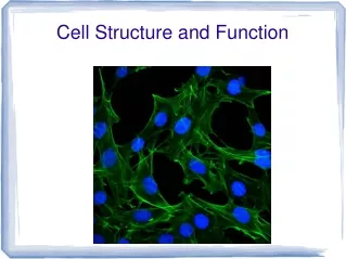
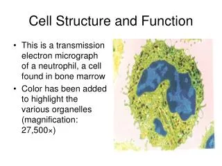
Cell Structure and Function. This is a transmission electron micrograph of a neutrophil, a cell found in bone marrow Color has been added to highlight the various organelles (magnification: 27,500×) . Life Is Cellular . Look closely at a part of a living thing, and what do you see?

Cell Structure and Function
E N D
Presentation Transcript
Cell Structure and Function • This is a transmission electron micrograph of a neutrophil, a cell found in bone marrow • Color has been added to highlight the various organelles (magnification: 27,500×)
Life Is Cellular • Look closely at a part of a living thing, and what do you see? • Hold a blade of grass up against the light, and you see tiny lines running the length of the blade • Examine the tip of your finger, and you see the ridges and valleys that make up fingerprints • Place an insect under a microscope, and you see the intricate structures of its wings and the spikes and bristles that protect its body • As interesting as these close-up views may be, however, they're only the beginning of the story • Look closer and deeper with a more powerful microscope, and you'll see that there is a common structure that makes up every living thing — the cell
The Discovery of the Cell • “Seeing is believing,” an old saying goes • It would be hard to find a better example of this than the discovery of the cell • Without the instruments to make them visible, cells remained out of sight and, therefore, out of mind for most of human history • All of this changed with a dramatic advance in technology — the invention of the microscope
Early Microscopes • It was not until the mid-1600s that scientists began to use microscopes to observe living things • In 1665, Englishman Robert Hooke used an early compound microscope to look at a thin slice of cork, a plant material • Under the microscope, cork seemed to be made of thousands of tiny, empty chambers • Hooke called these chambers “cells” because they reminded him of a monastery's tiny rooms, which were called cells • One of Hooke's illustrations of cells is shown in the figure to the right • The term cell is used in biology to this day • We now know, however, that cells are not empty but contain living matter
Cork Cells • Using an early microscope, Hooke made this drawing of cork cells • In Hooke's drawings, the cells look like empty chambers because he was looking at dead plant matter • Today, we know that living cells are made up of many structures
Early Microscopes • In Holland around the same time, Anton van Leeuwenhoek used a single-lensmicroscope to observe pond water and other things • To his amazement, the microscope revealed a fantastic world of tiny living organisms that seemed to be everywhere, even in the very water he and his neighbors drank
The Cell Theory • Soon, numerous observations made it clear that cells were the basic units of life • In 1838, German botanist Matthias Schleiden concluded that all plants were made of cells • The next year, German biologist Theodor Schwann stated that all animals were made of cells • In 1855, the German physician Rudolf Virchow concluded that new cells could be produced only from the division of existing cells • These discoveries, confirmed by other biologists, are summarized in the cell theory, a fundamental concept of biology
The Cell Theory • The cell theory states: • All living things are composed of cells • Cells are the basic units of structure and function in living things • New cells are produced from existing cells
Exploring the Cell • Following in the footsteps of Hooke, Virchow, and others, modern biologists still use microscopes to explore the cell • However, today's researchers use microscopes and techniques more powerful than the pioneers of biology could have imagined • Researchers can use fluorescent labels and light microscopy to follow molecules moving through the cell • Confocal light microscopy, which scans cells with a laser beam, makes it possible to build three-dimensional images of cells and their parts • High-resolution video technology makes it easy to produce movies of cells as they grow, divide, and develop
Exploring the Cell • These new technologies make it possible for researchers to study the structure and movement of living cells in great detail • Unfortunately, light itself limits the detail, or resolution, of images that can be made with the light microscope • Like all forms of radiation, light waves are diffracted, or scattered, as they pass through matter, making it impossible to visualize tiny structures such as proteins and viruses with light microscopy
Exploring the Cell • By contrast, as shown in the figure below, electron microscopes are capable of revealing details as much as 1000 times smaller than those visible in light microscopes because the wavelengths of electrons are much shorter than those of light • Transmission electron microscopes(TEMs) make it possible to explore cell structures and large protein molecules • Because beams of electrons can only pass through thin samples, cells and tissues must be cut first into ultrathin slices before they can be examined under a microscope
Exploring the Cell • With scanning electron microscopes (SEMs), a pencillike beam of electrons is scanned over the surface of a specimen • For SEM images, specimens do not have to be cut into thin slices to be visualized • The scanning electron microscope produces stunning three-dimensional images of cells • Because electrons are easily scattered by molecules in the air, samples examined in both types of electron microscopes must be placed in a vacuum in order to be studied • As a result, researchers chemically preserve their samples first and then carefully remove all of the water before placing them in the microscope • This means that electron microscopy can be used to visualize only nonliving, preserved cells and tissues
Exploring the Cell • In the 1990s, researchers perfected a new class of microscopes that produce images by tracing the surfaces of samples with a fine probe • These scanning probe microscopes have revolutionized the study of surfaces and made it possible to observe single atoms • Unlike electron microscopes, scanning probe microscopes can operate in ordinary air and can even show samples in solution • Researchers have already used scanning probe microscopes to image DNA and protein molecules as well as a number of important biological structures
Prokaryotes and Eukaryotes • Cells come in a great variety of shapes and an amazing range of sizes • Although typical cells range from 5 to 50 micrometers in diameter, the tiniest mycoplasma bacteria are only 0.2 micrometers across, so small that they are difficult to see under even the best light microscopes • In contrast, the giant amoeba Chaos chaos may be 1000 micrometers in diameter, large enough to be seen with the unaided eye as a tiny speck in pond water • Despite their differences, all cells have two characteristics in common • They are surrounded by a barrier called a cell membrane; and, at some point in their lives, they contain the molecule that carries biological information—DNA
Prokaryotes and Eukaryotes • Cells fall into two broad categories, depending on whether they contain a nucleus • The nucleus (plural: nuclei) is a large membrane-enclosed structure that contains the cell's genetic material in the form of DNA • A membrane is a thin layer of material that serves as a covering or lining • The nucleus controls many of the cell's activities • Eukaryotes are cells that contain nuclei • Prokaryotes are cells that do not contain nuclei • Both words derive from the Greek words karyon, meaning “kernel,” or nucleus, and eu, meaning “true,” or pro, meaning “before” • These words reflect the idea that prokaryotic cells evolved before nuclei developed
Prokaryotes • Prokaryotic cells are generally smaller and simpler than eukaryotic cells, although there are many exceptions to this rule • Prokaryotic cells have genetic material that is not contained in a nucleus • Some prokaryotes contain internal membranes, but prokaryotes are generally less complicated than eukaryotes: • NO MEMBRANE BOUND ORGANELLES • Despite their simplicity, prokaryotes carry out every activity associated with living things • They grow, reproduce, respond to the environment, and some can even move by gliding along surfaces or swimming through liquids • The organisms we call bacteria are prokaryotes
Eukaryotes • Eukaryotic cells are generally larger and more complex than prokaryotic cells • Eukaryotic cells generally contain dozens of structures and internal membranes, and many are highly specialized: • MEMBRANE BOUND ORGANELLES • Eukaryotic cells contain a nucleus in which their genetic material is separated from the rest of the cell • Eukaryotes display great variety • Some eukaryotes live solitary lives as single-celled organisms • Others form large, multicellular organisms. Plants, animals, fungi, and protists are eukaryotes
Cytoplasmic Organelles • Little organs • Specialized cellular compartments, each performing its own job to maintain the life of the cell • Membranous organelles: • Bounded by a membrane similar in composition to the plasma membranre (minus the glycocalyx) • This membrane enables them to maintain an internal environment different from that of the surrounding cytosol • Examples: • Mitochondria • Peroxisomes • Lysosomes • Endoplasmic reticulum • Golgi apparatus • Nonmembranous organelles: • Examples: • Cytoskeleton • Centrioles • Ribosomes
Eukaryotic Cell Structure • In some respects, the eukaryotic cell is like a factory • The first time you look at a microscope image of a cell, the cell seems impossibly complex • Look closely at a eukaryotic cell, however, and patterns begin to emerge • To see those patterns more clearly, we'll look at some structures that are common to eukaryotic cells, shown in the figure at right • Because many of these structures act as if they are specialized organs, these structures are known as organelles, literally “little organs”
Characteristics of Cells • All cells have the same basic parts and some common functions • A generalized human cell contains the plasma membrane, the cytoplasm, and the nucleus
Eukaryotic Cell Structure • Cell biologists divide the eukaryotic cell into two major parts: • Nucleus • Cytoplasm • The cytoplasm is the portion of the cell outside the nucleus • As you will see, the nucleus and cytoplasm work together in the business of life
Nucleus • In the same way that the main office controls a large factory, the nucleus is the control center of the cell • The nucleus contains nearly all the cell's DNA and with it the coded instructions for making proteins and other important molecules • The structure of the nucleus is shown in the figure at right
THE NUCLEUS • The nucleus is the control center of the cell and contains the cellular DNA • Most cells have only one nucleus, but very large cells may be multinucleate • Presence of more than one nucleus usually signifies that a larger-than-usual cytoplasmic mass must be regulated • All body cells except mature red blood cells (anucleate) have nuclei • The nucleus is larger than the cytoplasmic organelles • It has three regions: • Nuclear envelope (membrane) • Nucleoli • Chromatin
Nuclear Envelope • The nucleus is surrounded by a nuclear envelope composed of two membranes • The nuclear envelope is dotted with thousands of nuclear pores, which allow material to move into and out of the nucleus • Like messages, instructions, and blueprints moving in and out of a main office, a steady stream of proteins, RNA, and other molecules move through the nuclear pores to and from the rest of the cell
Nuclear Envelope • Is a double-membrane barrier (separated by a fluid-filled space) surrounding the nucleus • Outer membrane is continuous with the rough ER of the cytoplasm and is studded with ribosomes on its external face • Inner membrane is lined by a network of protein filaments ( the nuclear lamina) that maintains the shape of the nucleus • At various points, nuclear pores penetrate areas where the membranes of the nuclear envelope fuse • A complex of proteins, called a pore complex, lines each nuclear pore and regulates passage of large particles into and out of the nucleus • Like other cell membranes, the nuclear envelope is selectively permeable, but here passage of substances is much freer than elsewhere • Protein molecules imported from the cytoplasm and RNA molecules exported from the nucleus pass easily through the relatively large pores • The nuclear envelope encloses the fluid and solutes of the nucleus, the nucleoplasm
Chromatin • The granular material you can see in the nucleus is calledchromatin • Chromatin consists of DNA bound to protein • Most of the time, chromatin is spread throughout the nucleus • When a cell divides, however, chromatin condenses to form chromosomes • These distinct, threadlike structures contain the genetic information that is passed from one generation of cells to the next
Chromatin • (a): Appears as a fine, unevenly stained network, but special techniques reveal it as a system of bumpy threads weaving their way through the nucleoplasm • Is roughly half DNA, the genetic material of the cell, and half globular histone proteins: • Nucleosomes are the fundamental unit of chromatin, consisting of discus-shaped cores or clusters of eight histone proteins connected like beads on a string by a DNA molecule • DNA winds around each nucleosome and continues on to the next cluster via linker DNA segments
Chromatin • Histones provide physical means for packing the very long DNA molecules in a compact, orderly way, they also play an important role in gene regulation: • In a nondividing cell, addition of phosphate or methyl groups to histone exposes different DNA segments, or genes, so that they can dictate the specifications for protein synthesis • When a cell is preparing to divide, chromatin condenses into dense, rodlike chromosomes • Chromosome compactness avoids entanglement and breakage of the delicate chromatin strands during the movements that occur during celldivision
Nucleolus • Most nuclei also contain a small, dense region known as the nucleolus • The nucleolus is where the assembly of ribosomes begins
Nucleoli • Dark-staining spherical bodies within the nucleus • NOT membrane bound • There are typically one or two nucleoli per nucleus, but there may be more • Site of the assembly of ribosomal subunits: • Therefore, large in actively growing cells that are making large amounts of tissue proteins
NUCLEUS The nucleus is surrounded by a double membrane called the nuclear envelope. Inside the envelope is chromatin (combo of DNA and protein) which will become chromosomes. The nucleolus is the site where ribosomes are synthesized and partially assembled. The nucleus is porous and is the site where our genetic information is held.
Endoplasmic Reticulum • Eukaryotic cells also contain an internal membrane system known as the endoplasmic reticulum, or ER, as shown in the figure at right • The endoplasmic reticulum is the site where lipid components of the cell membrane are assembled, along with proteins and other materials that are exported from the cell
Ribosomes • One of the most important jobs carried out in the cellular “factory” is making proteins • Proteins are assembled on ribosomes • Ribosomesare small particles of RNA and protein found throughout the cytoplasm • They produce proteins by following coded instructions that come from the nucleus • Each ribosome, in its own way, is like a small machine in a factory, turning out proteins on orders that come from its “boss”—the cell nucleus • Cells that are active in protein synthesis are often packed with ribosomes
Ribosomes • (a):Small staining granules consisting of protein and ribosomal RNA • Each ribosome has two globular subunits that fit together • Site of protein synthesis
Ribosomes • Some float freely in the cytoplasm: • Make soluble proteins that function in the cytosol • Some are attached to membranes, forming a complex called the rough endoplasmic reticulum: • Synthesize proteins destined either for incorporation into cell membranes or for export from the cell • Ribosomes can switch back-and-forth between the two types
Endoplasmic Reticulum • The portion of the ER involved in the synthesis of proteins is called rough endoplasmic reticulum, or rough ER • It is given this name because of the ribosomes found on its surface • Newly made proteins leave these ribosomes and are inserted into the rough ER, where they may be chemically modified
Endoplasmic Reticulum • Proteins that are released, or exported, from the cell are synthesized on the rough ER, as are many membrane proteins • Rough ER is abundant in cells that produce large amounts of protein for export • Other cellular proteins are made on “free” ribosomes, which are not attached to membranes
Endoplasmic Reticulum • The other portion of the ER is known as smooth endoplasmic reticulum (smooth ER) because ribosomes are not found on its surface • In many cells, the smooth ER contains collections of enzymes that perform specialized tasks, including the synthesis of membrane lipids and the detoxification of drugs • Liver cells, which play a key role in detoxifying drugs, often contain large amounts of smooth ER
Endoplasmic reticulum • Is an extensive system of interconnected tubes and parallel membranes enclosing fluid-filled cavities, called cisternae, that coils and twist throughout the cytosol • Continuous with the nuclear membrane • Two varieties: • Rough ER • Smooth ER