Download
1 / 40
400 likes | 518 Vues
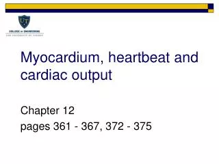
Myocardium, heartbeat and cardiac output. Chapter 12 pages 361 - 367, 372 - 375. Myocardium (cardiac muscle). Heart is myocardium lined with endothelial cells facing blood Myocardium combines properties of skeletal and smooth muscle

E N D
Myocardium, heartbeat and cardiac output Chapter 12 pages 361 - 367, 372 - 375
Myocardium (cardiac muscle) • Heart is myocardium lined with endothelial cells facing blood • Myocardium combines properties of skeletal and smooth muscle • Same sarcomere structure as skeletal muscle with light and dark bands • Fibers are shorter and have more branching when compared to skeletal muscle fibers • Excitation-contraction coupling is regulated by troponin and tropomyosin as in skeletal muscle • Smooth muscle myosin needs Ca2+-calmodulin complex to hydrolyze ATP
Figure 9-39 Join adjacent cardiac muscle cells Hold the cells together and myofibrils attach to
Myocardium (cardiac muscle) • Electrical conduction is similar to smooth muscle • No external innervation, neuromuscular junction, or ligand-gated AChRs required as in skeletal muscle • Some myocardial fibers undergo spontaneous pacemaker activity without external input • Many fibers are connected via gap junctions like smooth muscle to conduct pacemaker potentials
Myocardium (cardiac muscle) • Spontaneous activity and other contractile machinery modulated by hormones and neurotransmitters to alter cardiac output • Epinephrine/norepinephrine – b adrenergic receptors (GPCRs) • Acetylcholine – muscarinic AChRs (GPCRs) • Nicotinic AChRs are ligand-gated ion channels in neuromuscular junction
Which of the following are found in both cardiac and skeletal muscle fibers? • Gap junctions • Sarcomeres • Nicotinic AChRs • Muscarinic AChRs • b-adrenergic receptors
Electrical conduction in heart • Heart contains non-contractile myocardium for heartbeat coordination • Approximately 1% of myocardial fibers are non-contractile • Extensively connected by gap junctions • Remember that depolarization of one cell can be rapidly transmitted to a second cell via ion flow through gap junction channels • Spread wave of electrical excitation and coordinate contraction of contractile myocardium
Conducting system of the heart
Sequence of cardiac excitation QRS ventricles contract T ventricles repolarize P atria contract
Pacemaker potentials and myocardial excitability • Spontaneous APs in SA node do not require external depolarization • Peripheral nerves and skeletal muscle require synaptic transmission and EPSPs • SA node will spontaneously depolarize at rest without external excitation
Membrane potential from cardiac node cell Membrane potential from ventricular muscle cell
Pacemaker potentials and myocardial excitability • Unlike SA node, APs in contractile myocardium still require external source of depolarization • This external depolarization is provided by gap junctions • APs in contractile myocardium have much longer duration than skeletal muscle and peripheral nerves • Long AP functions to produced sustained contraction instead of a twitch
Excitation-contraction coupling • Some differences between cardiac and skeletal muscle excitation-contraction coupling • External Ca2+ required • Larger fraction of cytosolic Ca2+ comes from VGCCs (voltage gated calcium channels) that remain open during plateau AP
Excitation-contraction coupling • SR does not encircle cardiac A-M filament bundles • Not all A-M filaments available for cycling during cardiac AP • Strength of contraction modulated by increasing Ca2+ not by tetanus or recruiting fibers • Longer refractory period of cardiac muscle
Excitation-contraction Coupling in Cardiac Muscle L-type Ca channels means “long lasting current”, so this prolongs the AP and the twitch as compared to skeletal muscle
Timing of AP and twitch tension in Skeletal and Cardiac Muscle
Electrocardiogram • Electrocardiogram (ECG or EKG) records electrical signals produced by myocardial tissue due cardiac cycle • Coordinated contraction of heart muscle produces an large electrical signal due to bulk ion flow • Voltage changes due to bulk ion flow can be recorded with surface electrodes • EKG amplitude is proportional to V(t) and dV/dt • Characteristic waveform is observed and can be used to diagnose problems
EKG in Healthy Person & 2 Cases of AV Block Partial block, every other atrial impulse works Total block, ventricles driven by slow pacemaker cell in bundle of His
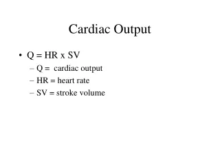
Cardiac output • The average flow rate of blood through heart per minute is known as cardiac output (CO) • CO is product of SV (stroke volume) and heart rate (HR) • CO = SV x HR • Can control heart rate and stroke volume to change cardiac output during rest or periods of intense activity • Heart rate change be modulated by altering excitability of pacemaker cells
Cardiac output • SV = EDV – ESV, can change SV in two ways • Systemic vasculature can control amount of blood returned to ventricle following diastole (EDV) • Ventricular contractility can control amount of blood left in ventricle following systole (ESV) • Control of CO is accomplished via autonomic nervous system • Parasympathetic system decreases CO • Sympathetic system increases CO
Control of heart rate • Normal heart will beat around 100 beats/min without any autonomic innervation • Numerous sympathetic and parasympathetic efferents terminate in SA node • At rest, parasympathetic system is more active so resting HR is 70 beats/min, below inherent rate of 100 beats/min
Control of heart rate • Sympathetic system increases HR by increasing inward cation (If) and low-threshold T-type Ca2+ currents in SA node • Larger inward current increases rate of spontaneous depolarization • Parasympathetic fibers decrease HR by decreasing If and T-type Ca2+ currents • Parasympathetic fibers also increase K+ channel activity • Smaller inward current reduces rate of spontaneous depolarization
Effects of sympathetic and parasympathetic nerves on SA nodal cell
GPCRs and heart rate • Activation of GPCRs will effect contractility and heart rate via modulation of voltage-gated ion channels • Sympathetic increase of heart rate • NE → b adrenergic GPCR → activates adenylyl cyclase → increases cAMP → activates PKA → increased phosphorylation of If and T-type channels → more inward current → increased dVm/dt • Phosphorylated channels are more likely to open at “resting” Vm • Parasympathetic decrease of heart rate • ACh → mAChR GPCR → inhibits adenylyl cyclase → decreases cAMP → deactivates PKA → decreased phosphorylation of If and T-type channels → less inward current → decreased dVm/dt • Dephosphorylated channels are less likely to open at “resting” Vm
Control of stroke volume – ventricular filling • Changes in EDV and ESV affect SV • Remember SV = EDV – ESV • EDV alters ability of ventricles to eject blood • Relationship between SV and EDV is called Frank-Starling mechanism • Consequence of relationship between muscle fiber length and tension • Stretched cardiac muscle generates more tension
Ventricular Function Curve, EDV is major determinant of ventricular stretch which yields more forceful contraction
Control of stroke volume – ventricular filling • Therefore filling ventricles more will increase ability of ventricles to eject blood • Direct relationship venous return → atrial volume → ventricular filling → EDV → SV • Main function is to make sure pulmonary and systemic blood flows are equal • Prevents accumulation of blood in pulmonary or systemic vessels
Control of stroke volume - afterload • Increased arterial pressure increases ESV (end systolic volume) • Increased arterial pressure will shorten ventricular ejection • Valve closes when Parterial > Pventricle • Less time to eject blood from ventricle during single cycle, so stroke volume decreases
Control of stroke volume - afterload • If blood pressure is elevated, heart must work harder to eject same amount of fluid • This requires increased ventricular contractility • Arteries can control systemic pressures via constriction and dilation • Preload – venous return that fills ventricle to increase SV • Afterload – arterial pressure that heart must work against or result is decrease in SV • Changes in SV due to preload and afterload are “passive” effects
Control of stroke volume - contractility • EDV is somewhat affected by atrial contractility • ESV is controlled by ventricular contractility • Contractility changes are “active”, affected by sympathetic and parasympathetic innervation • Reflects increased myocardial activity independent of length-tension relation
Sympathetic stimulation causes increased contractility of ventricular muscle
Rate of force development and relaxation increase as well as the force developed
Control of stroke volume - contractility • Main target is control of Ca2+ influx through VGCCs (voltage gated calcium channels) and CICR (calcium induced calcium release) • Atrial myocardium has sympathetic and parasympathetic innervation • mAChRs and b adrenergic GPCRs • Ventricular myocardium only has sympathetic innervation • b adrenergic GPCRs
Mechanisms of sympathetic effects on cardiac muscle contractility
Major factors determining cardiac output. Reversal of arrows illustrate how cardiac output is decreased