Download
1 / 22
220 likes | 396 Vues
Genes are composed of nucleic acids (usually DNA). Pneumococcus can be transformed from an avirulent to a virulent strain DNA is the transforming principle DNA in bacteriophage particles appears in the progeny, but very little protein does. Structures of nucleic acids. Nucleotides

E N D
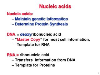
Genes are composed of nucleic acids (usually DNA) • Pneumococcus can be transformed from an avirulent to a virulent strain • DNA is the transforming principle • DNA in bacteriophage particles appears in the progeny, but very little protein does.
Structures of nucleic acids Nucleotides DNA structures Sedimentation and Electrophoresis
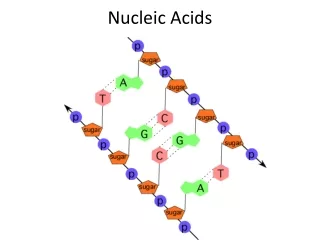
A simple view of DNA AGCCTCGCAT TCGGAGCGTA
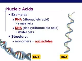
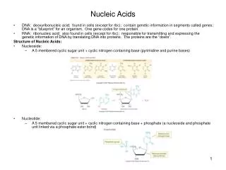
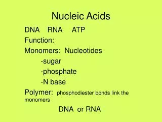
Nucleotides • 3 components to nucleotides: • Purine or pyrimidine base • Ribose (RNA) or 2-deoxyribose (DNA) sugar • Phosphate • Base + sugar = Nucleoside • Base + sugar + phosphate = Nucleotide
Types of bases in nucleotides Pyrimidine Amino- Keto-
Nucleotides: purine bases 6-aminopurine A keto-purine
Bases are attached to C1’ of the sugar via an N-glycosidic bond 2’-deoxy- , a nucleoside
1st phosphate is a phosphoester, others are attached as phosphoanhydrides. Phosphate is attached to C5’ of the sugar g b a
Structure of a dinucleotide The 3’ C of one nucleotide is linked to the 5’ C of the next nucleotide in a phosphodiester linkage.
Nucleic acids are linear chains of nucleotides • The 3’ C of one nucleotide is linked to the 5’ C of the next nucleotide. • The linkage is by a phosphoester. • The chain has an orientation defined by the sugar-phosphage backbone. • One terminal nucleotide has a “free” 5’ end, and the other has a “free” 3’ end. • Thus we designate orientation by 5’ to 3’.
More on orientation of chains of nucleic acids • 5’ ACTG 3’ is different from 3’ ACTG 5’ • Unless specified otherwise, a chain is written with the 5’ end on the left and the 3’ end on the right. • When complementary strands in DNA are written, usually the top strand is written 5’ to 3’, left to right, and the bottom strand is written 3’ to 5’, left to right. 5’ GATTCGTACCG 3’ CTAAGCATGGC
Basics of DNA structure • 2 complementary strands of nucleic acids • Complementarity is based on H-bonding between • Keto bases with amino bases • Pyrimidines with purines • A pairs with T (or U) • G pairs with C • The complementary strands are antiparallel. • The complementary strands are coiled around each other.
Duplex DNA • Two strands coil around each other. • Right-handed coils (B form and A. • Coils form major and minor grooves. • Strands have opposite polarity (antiparallel). • Opposing bases in strands are complementary. • Different edges of paired bases are exposed in major and minor grooves. • Sugar-phosphate backbone is on the outside, bases on the inside • B-form DNA: base pairs are close to center of long axis of the duplex. • A-form nucleic acids: base pairs stack away from long axis.
Implications of complementarity • One chain (strand) of DNA can serve as the template for synthesis of the complementary chain. • DNA replication: sequence of nucleotides in one chain of the duplex determines the sequence of nucleotides in the other chain. • Transcription: sequence of nucleotides in one chain of the duplex determines the sequence of nucleotides in mRNA or its precursor.
Base pairs in DNA Major groove Major groove Minor groove Minor groove Guanine : Cytosine Adenine : Thymine
Major types of duplex nucleic acid structures • B form • Most common form of DNA • Right handed DNA-DNA helix • Base pairs stack close to DNA central axis • A form • right handed RNA-DNA and RNA-RNA helix • Base pairs stack away from the central axis • Z form DNA • Repeating purines and pyrimidines • Left-handed helix • May serve as some regulatory signal in cells
Forms of nucleic acid duplexes A-form (e.g. duplex RNA) Z DNA B-form DNA
Helical parameters for B, A and Z nucleic acids BAZ helix sense RH RH LH bp per turn 10 11 12 vertical rise per bp 3.4 2.56 3.7 Angstroms rotation per bp +36 +33 -30 degrees helical diameter 19 23 18 Angstroms
Factors that affect melting temperature, p. 85 • The melting tempera-ture (Tm) increases as • Increase G+C • Increase ionic strength (or m) • Tm decreases as • Increase denaturants • Increase number of mismatches Tm = 0.41 (% G+C) + 16.6 log M + 81.5 -0.7 (% formamide) -1o (% mismatch)
Example of gel electrophoresis Markers Alpha-globin gene PCR product 217 bp 400 base pairs 300 200 100