Download
1 / 60
630 likes | 934 Vues
Chapter 18 The Circulatory System: Blood. The Circulatory System: Blood. Introduction Erythrocytes Blood types Leukocytes Platelets Hemostasis – the control of bleeding. Functions of Circulatory System. Transport O 2 , CO 2 , nutrients, wastes, hormones, and heat Protection

E N D
Chapter 18 The Circulatory System: Blood
The Circulatory System: Blood • Introduction • Erythrocytes • Blood types • Leukocytes • Platelets • Hemostasis – the control of bleeding
Functions of Circulatory System • Transport • O2, CO2, nutrients, wastes, hormones, and heat • Protection • WBCs, antibodies, and platelets • Regulation • fluid regulation and buffering
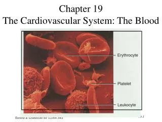
Blood • Adults have 4-6 L of blood • plasma, a clear extracellular fluid • formed elements (blood cells and platelets) • Centrifuge blood to separate components
Properties of Blood • Viscosity - resistance to flow • whole blood 5 times as viscous as water • Osmolarity • total molarity of dissolved particles • sodium ions, protein, and RBCs • high osmolarity • causes fluid absorption into blood, raises BP • low osmolarity • causes fluid to remain in tissues, may result in edema
Plasma and Plasma Proteins • Plasma – liquid portion of blood • serum remains after plasma clots • 3 major categories of plasma proteins • albumins - most abundant • contributes to viscosity and osmolarity, influences blood pressure, flow and fluid balance • globulins (antibodies) • provide immune system functions • alpha, beta and gamma globulins • fibrinogen • precursor of fibrin threads that help form blood clots • Plasma proteins formed by liver • except globulins (produced by plasma cells)
Nonprotein Components of Plasma • Nitrogenous compounds • amino acids • from dietary protein or tissue breakdown • nitrogenous wastes (urea) • toxic end products of catabolism • normally removed by the kidneys • Nutrients • glucose, vitamins, fats, minerals, etc • O2 and CO2 • Electrolytes • Na+ makes up 90% of plasma cations
Nutritional Needs for Erythropoiesis • Vitamin B12 and folic acid • rapid cell division • Vitamin C and copper • cofactors for enzymes synthesizing RBCs
Erythrocytes (RBCs) • Disc-shaped cell with thick rim • 7.5 M diameter and 2.0 m thick at rim • blood type determined by surface glycoprotein and glycolipids • cytoskeletal proteins give membrane durability
Erythrocytes (RBCs) Function • Gas transport - major function • increased surface area/volume ratio • due to loss of organelles during maturation • increases diffusion rate of substances • 33% of cytoplasm is hemoglobin (Hb) • O2 delivery to tissue and CO2 transport to lungs • Carbonic anhydrase (CAH) • produces carbonic acid from CO2 and water • important role in gas transport and pH balance
Hemoglobin (Hb) Structure • Heme groups • conjugate with each protein chain • hemoglobin molecule can carry four O2 • binds oxygen to ferrous ion (Fe2+) • Globins - 4 protein chains • 2 alpha and 2 beta chains • fetal Hb - gamma replace beta chains; binds O2 better
Erythrocytes and Hemoglobin • RBC count and hemoglobin concentration indicate amount of O2 blood can carry • hematocrit (packed cell volume) - % of blood composed of cells • men 42- 52% cells; women 37- 48% cells • hemoglobin concentration of whole blood • men 13-18g/dL; women 12-16g/dL • RBC count • men 4.6-6.2 million/L; women 4-2-5.4 million/L • Values are lower in women • androgens stimulate RBC production • women have periodic menstrual losses
Hemopoiesis • Adult produces 400 billion platelets, 200 billion RBCs and 10 billion WBCs every day • Hemopoietic tissues produce blood cells • yolk sac produces stem cells • colonize fetal bone marrow, liver, spleen and thymus • liver stops producing blood cells at birth • spleen remains involved with WBC production • lymphoid hemopoiesis occurs in widely distributed lymphoid tissues (thymus, tonsils, lymph nodes, spleen and peyers patches in intestines) • red bone marrow • pluripotent stem cells • myeloid hemopoiesis produces RBCs, WBCs and platelets
Erythrocyte Homeostasis • Negative feedback control • drop in RBC count causes kidney hypoxemia • EPO production stimulates bone marrow • RBC count in 3 - 4 days • Stimulus for erythropoiesis • low levels O2 • increase in exercise • loss of lung tissue in emphysema
Nutritional Needs for Erythropoiesis • Iron - key nutritional requirement • lost daily through urine, feces, and bleeding • men 0.9 mg/day and women 1.7 mg/day • low absorption requires consumption of 5-20 mg/day • dietary iron: ferric (Fe3+) and ferrous (Fe2+) • stomach acid converts Fe3+ to absorbable Fe2+ • gastroferritin binds Fe2+ and transports it to intestine • absorbed into blood and binds to transferrin for transport • bone marrow for hemoglobin, muscle for myoglobin and all cells use for cytochromes in mitochondria • liver apoferritin binds to create ferritin for storage
Erythrocyte Production • 2.5 million RBCs/sec • Development takes 3-5 days • reduction in cell size, increase in cell number, synthesis of hemoglobin and loss of nucleus • First committed cell - erythrocyte colony forming unit • has receptors for erythropoietin (EPO) from kidneys • Erythroblasts multiply and synthesize hemoglobin • Discard nucleus to form a reticulocyte • named for fine network of endoplasmic reticulum • 0.5 to 1.5% of circulating RBCs
Erythrocytes Recycle/Disposal • RBCs lyse in narrow channels in spleen • Macrophages in spleen • digest membrane bits • separate heme from globin • globins hydrolyzed into amino acids • iron removed from heme • heme pigment converted to biliverdin (green) • biliverdin converted to bilirubin (yellow) • released into blood plasma (kidneys - yellow urine) • liver secretes into bile • concentrated in gall bladder: released into small intestine; bacteria create urobilinogen (brown feces)
Erythrocyte Disorders • Polycythemia - an excess of RBCs • primary polycythemia • cancer of erythropoietic cell line in red bone marrow • RBC count as high as 11 million/L; hematocrit 80% • secondary polycythemia • from dehydration, emphysema, high altitude, or physical conditioning • RBC count up to 8 million/L • Dangers of polycythemia • increased blood volume, pressure, viscosity • can lead to embolism, stroke or heart failure
Anemia - Causes • Inadequate erythropoiesis or hemoglobin synthesis • inadequate vitamin B12 from poor nutrition or lack of intrinsic factor (pernicious anemia) • iron-deficiency anemia • kidney failure and insufficient erythropoietin • aplastic anemia - complete cessation • Hemorrhagic anemias • Hemolytic anemias
Anemia - Effects • Tissue hypoxia and necrosis (short of breath and lethargic) • Low blood osmolarity (tissue edema) • Low blood viscosity (heart races and pressure drops)
Sickle-Cell Disease • Hereditary Hb ‘defect’ of African Americans • recessive allele modifies hemoglobin structure • sickle-cell trait - heterozygous for HbS • individual has resistance to malaria • HbS indigestible to malaria parasites • sickle-cell disease - homozygous for HbS • individual has shortened life • in low O2 concentrations HbS causes cell elongation and sickle shape • cell stickiness causes agglutination and blocked vessels • intense pain; kidney and heart failure; paralysis; stroke • chronic hypoxemia reactivates hemopoietic tissue • enlarging spleen and bones of cranium
Sickle-Cell Diseased Erythrocyte Fig. 18.10
Antigens and Antibodies • Antigens • unique molecules on cell surface • used to distinguish self from foreign • foreign antigensgenerate immune response • Antibodies • secreted by plasma cells • as part of immune response to foreign matter • Agglutination • antibody molecule binding to antigens • causes clumping
Blood Types • RBC antigens • agglutinogens; A and B • on RBC surface
ABO Group • Your ABO blood type is determined by presence or absence of antigens (agglutinogens) on RBCs • type A person has A antigens • type B person has B antigens • type AB has both antigens • type O has neither antigen • most common - type O • rarest - type AB
Plasma antibodies • Antibodies (agglutinins); anti-A and -B • Appear 2-8 months after birth; at maximum concentration at 10 yr. • Anti -A and/or -B (both or none) are in plasma • you do not form antibodies against your antigens • Agglutination • each antibody can attach to several foreign antigens at the same time • Responsible for mismatched transfusion reaction
Transfusion Reaction • Agglutinated RBCs block blood vessels and hemolyze • free Hb blocks kidney tubules, causes death
Universal Donors and Recipients • Universal donor • Type O • lacks RBC antigens • donor’s plasma may have antibodies against recipient’s RBCs • may give packed cells (minimal plasma) • Universal recipient • Type AB • lacks plasma antibodies; no anti- A or B
Rh Group • Rh (D) agglutinogens discovered in rhesus monkey in 1940 • Rh+ blood type has D agglutinogens on RBCs • Rh frequencies vary among ethnic groups • Anti-D agglutinins not normally present • form in Rh- individuals exposed to Rh+ blood • Rh- woman with an Rh+ fetus or transfusion of Rh+ blood • no problems with first transfusion or pregnancy
Hemolytic Disease of Newborn • Occurs if mother has formed antibodies and is pregnant with 2nd Rh+ child • Anti-D antibodies can cross placenta • Prevention • RhoGAM given to pregnant Rh- women • binds fetal agglutinogens in her blood so she will not form Anti-D antibodies
Hemolytic Disease of Newborn • Rh antibodies attack fetal blood • causing severe anemia and toxic brain syndrome Fig. 18.16
Leukocytes (WBCs) • 5,000 to 10,000 WBCs/L • Conspicuous nucleus • Travel in blood before migrating to connective tissue • Protect against pathogens
Leukocyte Descriptions • Granulocytes • neutrophils (60-70%) • fine granules in cytoplasm; 3 to 5 lobed nucleus • eosinophils (2-4%) • large rosy-orange granules; bilobed nucleus • basophils (<1%) • large, abundant, violet granules (obscure a large S-shaped nucleus) • Agranulocytes • lymphocytes (25-33%) • variable amounts of bluish cytoplasm (scanty to abundant); ovoid/round, uniform dark violet nucleus • monocytes (3-8%) • largest WBC; ovoid, kidney-, or horseshoe- shaped nucleus
Granulocyte Functions • Neutrophils ( in bacterial infections) • phagocytosis of bacteria • release antimicrobial chemicals • Eosinophils ( in parasitic infections or allergies) • phagocytosis of antigen-antibody complexes, allergens and inflammatory chemicals • release enzymes to destroy parasites • Basophils ( in chicken pox, sinusitis, diabetes) • secrete histamine (vasodilator) • secrete heparin (anticoagulant)
Agranulocyte Functions • Lymphocytes ( in diverse infections and immune responses) • destroy cells (cancer, foreign, and virally infected cells) • “present” antigens to activate other immune cells • coordinate actions of other immune cells • secrete antibodies and provide immune memory • Monocytes ( in viral infections and inflammation) • differentiate into macrophages • phagocytize pathogens and debris • “present” antigens to activate other immune cells
Complete Blood Count • Hematocrit • Hemoglobin concentration • Total count for RBCs, reticulocytes, WBCs, and platelets • Differential WBC count • RBC size and hemoglobin concentration per RBC
Leukocyte Life Cycle • Leukopoiesis • pluripotent stem cells – • myeloblasts – form neutrophils, eosinophils, basophils • monoblasts form monocytes • lymphoblasts form B and T lymphocytes and NK cells • T lymphocytes complete development in thymus • Red bone marrow stores and releases granulocytes and monocytes • Circulating WBCs do not stay in bloodstream • granulocytes leave in 8 hours and live 5 days longer • monocytes leave in 20 hours, transform into macrophages and live for several years • WBCs provide long-term immunity (decades)
Leukopoiesis Fig. 18.18
Leukocyte Disorders • Leukopenia - low WBC count (<5000/L) • causes: radiation, poisons, infectious disease • effects: elevated risk of infection • Leukocytosis = high WBC count (>10,000/L) • causes: infection, allergy and disease • differential count - distinguishes % of each cell type • Leukemia = cancer of hemopoietic tissue • myeloid and lymphoid - uncontrolled WBC production • acute and chronic - death in months or 3 years • effects - normal cell % disrupted; impaired clotting
Platelets • Small fragments of megakaryocyte cytoplasm • 2-4 m diameter; contain “granules” • amoeboid movement and phagocytosis • Normal Count - 130,000 to 400,000 platelets/L • Functions • secrete clotting factors and growth factors for vessel repair • initiate formation of clot-dissolving enzyme • phagocytize bacteria • chemically attract neutrophils and monocytes to sites of inflammation
Platelet Production -Thrombopoiesis • Stem cells (that develop receptors for thrombopoietin) become megakaryoblasts • Megakaryoblasts • repeatedly replicate DNA without dividing cytoplasm • forms gigantic cell called megakaryocyte (100 m in diameter, remains in bone marrow) • Megakaryocyte • infoldings of cytoplasm splits off cell fragments that enter bloodstream as platelets (live for 10 days) • some stored in spleen
Hemostasis • All 3 pathways involve platelets
Hemostasis - Vascular Spasm • Causes • pain receptors • some directly innervate constrictors • smooth muscle injury • platelets release serotonin (vasoconstrictor) • Effects • prompt constriction of a broken vessel • pain receptors - short duration (minutes) • smooth muscle injury - longer duration • provides time for other two clotting pathways
Hemostasis -Platelet Plug Formation • Endothelium smooth, coated with prostacyclin • Platelet plug formation • broken vessel exposes collagen • platelet pseudopods stick to damaged vessel and other platelets - pseudopods contract and draw walls of vessel together forming a platelet plug • platelets degranulate releasing a variety of substances • serotonin is a vasoconstrictor • ADP attracts and degranulates more platelets • thromboxane A2, an eicosanoid, promotes aggregation, degranulation and vasoconstriction • positive feedback cycle is active until break in vessel is sealed