Functions of the kidney
410 likes | 1.22k Vues
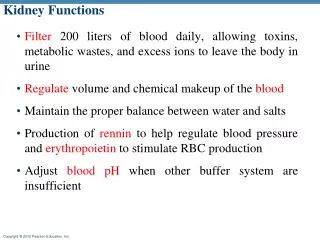
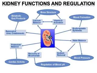
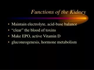
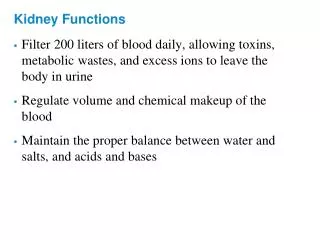
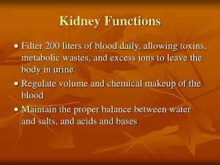
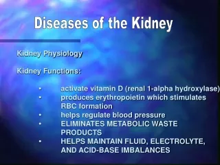
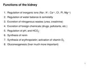
Functions of the kidney. 1. Regulation of inorganic ions (Na + , K + , Ca ++ , Cl - , Pi, Mg ++ ) 2. Regulation of water balance & osmolality 3. Excretion of nitrogenous wastes (urea, creatinine) 4. Excretion of foreign chemicals (drugs, pollutants, etc.) 5. Regulation of pH, and HCO 3 -

Functions of the kidney
E N D
Presentation Transcript
Functions of the kidney 1. Regulation of inorganic ions (Na+, K+, Ca++, Cl-, Pi, Mg++) 2. Regulation of water balance & osmolality 3. Excretion of nitrogenous wastes (urea, creatinine) 4. Excretion of foreign chemicals (drugs, pollutants, etc.) 5. Regulation of pH, and HCO3- 6. Synthesis of renin 7. Synthesis of erythropoietin; activation of vitamin D3 8. Gluconeogenesis (liver much more important)
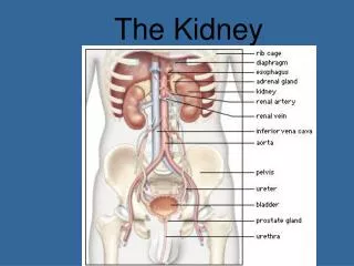
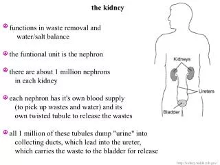
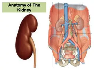
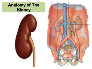
Structure of urinary system fig 14-1
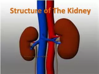
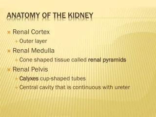
Structure of kidney fig 14-4
Structure of nephron & blood supply Nephron: Bowman’s capsule (C) proximal tubule (C) loop of Henle (M) distal tubule (C) collecting duct (C, M) Blood supply afferent arteriole (C) glomerular capillaries (C) efferent arteriole (C) peritubular capillaries (C) vasa recta (M) venule (C) (C) = cortex (M) = medulla fig 14-2
Structure of glomerulus (renal corpuscle) Blood flow: afferent arteriole glomerular capillaries efferent arteriole Filtration: from glomerular capillaries into Bowman’s capsule Cell types: juxtaglomerular apparatus (macula densa + juxtaglomerular cells) podocytes fig 14-3
Direction of filtration From plasma through capillary endothelial cell fenestrae & podocyte filtration slits into Bowman’s capsule. Fluid in Bowman’s capsule is protein-free filtrate of plasma fig 14-3b
Juxtaglomerular apparatus Macula densa: specialized cells in wall of distal tubule Juxtaglomerular cells: contain renin, sympathetic nerves fig 14-5
Filtration, reabsorption, secretion, excretion Filtration: glomerular capillaries Bowman’s capsule Secretion: peritubular capillaries tubular fluid Reabsorption: tubular fluid peritubular capillaries Excretion = filtration + secretion – reabsorption fig 14-6
Kidney handling of various substances Substance X: filtered & entirely secreted (rare) Substance Y: filtered & partially reabsorbed (Na+, K+, water) Substance Z: filtered & entirely reabsorbed (glucose, amino acids) fig 14-7
Glomerular filtration barrier fig 14-3c
Forces of filtration Compare with Starling forces in muscle capillaries fig 14-8
Important numbers Resting cardiac output: ~5L/min Renal blood flow: ~1.2 L/min (i.e. about 25% of CO) Renal plasma flow: ~650 ml/min (~55% of blood is plasma) Glomerular filtration rate: ~120 ml/min (i.e. about 20% of renal plasma flow) Therefore, of plasma flowing through glomerular capillaries, ~20% is filtered into Bowman’s capsule and only 80% enters the efferent arteriole GFR of 120 ml/min = ~180 L/day (i.e. plasma is filtered 60x each day) Urine flow rate = ~1 ml/min (i.e. 99+% water reabsorbed) fig 14-8
Reabsorption reabsorption fig 14-10
Reabsorption of glucose & amino acids Glucose & amino acids are freely filtered At normal plasma concentrations they are all entirely reabsorbed Hence, urine [glucose] & [amino acid] are zero (see substance Z fig 14-7) Mechanism: Na+ linked co-transport at the luminal membrane of the proximal tubule In an untreated diabetic, plasma [glucose] is high, therefore the amount of glucose filtered is greater then the maximum transport rate of the transporters; hence glucose appears in the urine.
Reabsorption of urea Urea is freely filtered As water is reabsorbed, the tubular [urea] rises Urea is passively reabsorbed down its concentration gradient (see substance Y fig 14-7) About half the filtered urea is excreted
Measurement of glomerular filtration rate Inulin (~5000 M.Wt. polysaccharide) prepared from plants Inulin is filtered, not reabsorbed, not secreted Therefore all the inulin that is filtered is excreted Inulin filtered = GFR x [inulin]plasma Inulin excreted = urine flow rate x [inulin]urine Therefore GFR = urine flow rate x [inulin]urine [inulin]plasma Clinically, creatinine is used to measure GFR Creatinine is released at a constant rate from muscle Creatinine properties are similar, but not identical, to inulin
Sodium balance Most NaCl intake added during food preparation Sweat output depends on body temperature Urine output of NaCl is regulated by blood pressure
Water balance Metabolically produced by oxidation of H-containing nutrients Insensible loss: expiration of 37 saturated air, evaporation through skin (different from sweat) Urine output regulated by vasopressin (antidiuretic hormone ADH)
Total body NaCl and extracellular volume total body NaCl extracellular osmolality vasopressin release • water retention by kidneys • • extracellular volume Because vasopressin release is sensitive to changes in osmolality, any change in total body NaCl will result in a proportional change in extracellular volume
Sodium handling by the kidney Of the sodium filtered ~99.5% is reabsorbed, 0.5% excreted Sympathetic nervous system regulates glomerular filtration rate Sodium reabsorption: ~70% from proximal tubule (unregulated) ~20% from ascending limb of loop of Henle (unregulated) ~5% from distal tubule (unregulated) 3-5% from collecting duct (regulated by aldosterone & atrial natriuretic peptide-less important)
Sympathetic nervous system on sodium excretion • 1. action on glomerular filtration rate • blood pressure discharge from carotid sinus SNS activity glomerular filtration rate Na+ filtered Na+ excreted • action on renin release • blood pressure afferent arteriole pressure renin • angiotensin I,II aldosterone Na+ reabsorbed • Na+ excreted
Renin angiotensin aldosterone system fig 14-19
Renin angiotensin aldosterone system on Na+ excretion fig 14-20
Renin angiotensin aldosterone system on Na+ excretion • Renin release stimulated by: • 1. sympathetic nervous activity • 2. blood pressure in afferent arteriole • Na+ and Cl- in tubular fluid at macula densa • Angiotensin II actions: • general vasoconstriction • stimulates aldosterone release from adrenal cortex • Aldosterone release stimulated by: • 1. plasma angiotensin II levels • 2. plasma potassium concentration
Actions of aldosterone fig 14-13 Aldosterone actions: Na+ channel activity, K+ channel activity, Na+/K+ ATPase pump Note: large Na+, K+ shows high concentration & vice versa
Atrial natriuretic peptide on Na+ excretion • ANP actions: • Na+ reabsorption from deep medullary collecting duct • 2. glomerular filtration rate • Both actions Na+ excretion fig 14-21
Water transport & vasopressin (ADH) dependence Transport mechanism: passive diffusion through aquaporin channels down osmotic gradient Reabsorption: ~99% of filtered water is reabsorbed Sites of reabsorption: ~70% from proximal tubule ~15% from descending limb of loop of Henle 0% from Henle’s ascending limb & distal tubule 0-15% from collecting duct depending on plasma vasopressin level
Vasopressin (ADH) release & actions • Vasopressin release stimulated by: • slight (1%) increase in plasma osmolality • large (~15%) reduction in plasma volume • Vasopressin action: • increases permeability of collecting duct to water • Renal medulla • has osmotic gradient from 300 mOsm/kg at cortical border to 1200 mOsm/kg at deepest part of medulla (mechanism not necessary) • ADH levels collecting duct permeability water reabsorption urine volume with osmolality
Vasopressin action fig 14-15
Water transport & vasopressin actions fig 14-23 fig 14-22
Sweating without water replacement fig 14-24 Sweat is hypotonic (i.e. osmolality < plasma) Gatorade story
Regulation of thirst • Sensation of thirst stimulated by: • 1% osmolality • 2. >15% blood volume • angiotensin II • 4. dry mouth, throat • Sensation of thirst inhibited by: • 1. GI metering of water intake fig 14-25
Potassium excretion Reabsorption from: proximal tubule Henle’s ascending limb (collecting duct) Secretion into: collecting duct (regulated process) fig 14-26
Regulation of potassium excretion fig 14-27