Download
1 / 24
270 likes | 729 Vues
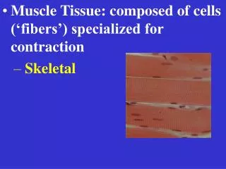
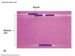
Figure 6.4b Muscle cells. Figure 6.1a Movement of bones. Figure 6.1b Movement of bones. Figure 6.3 Muscle structure. Epimysium. Bone. Tendon. Blood vessel. Fascicle (wrapped by perimysium). Endomysium (between individual muscle fibers). Muscle fiber.

E N D
Epimysium Bone Tendon Blood vessel Fascicle (wrapped by perimysium) Endomysium (between individual muscle fibers) Muscle fiber Figure 9.1a Connective tissue sheaths of skeletal muscle: epimysium, perimysium, and endomysium.
Sarcolemma Mitochondrion Myofibril Dark A band Light I band Nucleus (b) Diagram of part of a muscle fiber showing the myofibrils. Onemyofibril is extended afrom the cut end of the fiber. Figure 9.2b Microscopic anatomy of a skeletal muscle fiber.
Myosin filament Actin filament I band thin filaments only H zone thick filaments only M line thick filaments linked by accessory proteins Outer edge of A band thick and thin filaments overlap (e) Cross-sectional view of a sarcomere cut through in different locations. Figure 9.2e Microscopic anatomy of a skeletal muscle fiber.
Thin filament Actin Ca2+ ADP Myosin head P i Thick filament Myosin 1 Cross bridge formation. ADP ADP ATP hydrolysis P P i i The power (working) stroke. 2 4 Cocking of myosin head. ATP ATP 3 Cross bridge detachment. Figure 9.12 Cross Bridge Cycle
Thin filament (actin) Myosin heads Thick filament (myosin) Figure 9.4 Transmission electron micrograph of part of a sarcomere clearly showing the myosin heads forming cross bridges that generate the contractile force.
Axon terminal of neuromuscular junction Myelinated axon of motor neuron Action potential (AP) Sarcolemma of the muscle fiber Nucleus Figure 9.8 Events at the Neuromuscular Junction (1 of 4)
Figure 9.8 Events at the Neuromuscular Junction (2 of 4) Acetylcholine, a neurotransmitter, diffuses across the synaptic cleft and binds to receptors in the sarcolemma. Ca2+ Ca2+ Synaptic vesicle containing ACh Axon terminal of motor neuron Synaptic cleft Fusing synaptic vesicles ACh Sarcoplasm of muscle fiber
ACh binding opens ion channels that allow simultaneous passage of Na+ into the muscle fiber and K+ out of the muscle fiber. 5 K+ Na+ Postsynaptic mem- brane ion channel opens; ions pass. Figure 9.8 Events at the Neuromuscular Junction (3 of 4)
Resting Potential of muscle cells – • Potassium higher inside • Sodium higher outside • A voltage difference of about 90 mvolts • Action Potential – • A wave of depolarization that propogates from the point of stimulation over the entire membrane, followed by a wave of repolarization.
Axon terminal Open Na+ Channel Closed K+ Channel Na+ Synaptic cleft ACh– K+ Na+ K+ ACh Na+ K+ Generation and propagation of the action potential (AP) 2 Local depolarization: Na+ 1 Sarcoplasm of muscle fiber K+ Repolarization 3 Figure 9.9 Summary of events in the generation and propagation of an action potential in a skeletal muscle fiber.
Figure 9.5 Relationship of the sarcoplasmic reticulum and T tubules to myofibrils of skeletal muscle. Sarcolemma Triad T tubule Terminal cisternae of SR Myofibrils Mitochondria
Action potential is propagated along the sarcolemma and down the T tubules. 1 Steps in E-C Coupling: Sarcolemma Voltage-sensitive tubule protein T tubule Ca2+ release channel 2 Calcium ions are released. Terminal cisterna of SR Ca2+ Figure 9.11 Excitation-Contraction Coupling (3 of 4)