Download
1 / 43
460 likes | 679 Vues
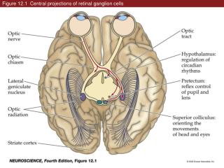
Figure 12.1 Central projections of retinal ganglion cells. Figure 12.2 The circuitry responsible for the pupillary light reflex. Figure 12.2 The circuitry responsible for the pupillary light reflex (Part 1). Figure 12.2 The circuitry responsible for the pupillary light reflex (Part 2).

E N D
Figure 12.2 The circuitry responsible for the pupillary light reflex
Figure 12.2 The circuitry responsible for the pupillary light reflex (Part 1)
Figure 12.2 The circuitry responsible for the pupillary light reflex (Part 2)
Figure 12.3 Projection of the visual fields onto the left and right retinas
Figure 12.3 Projection of the visual fields onto the left and right retinas (Part 1)
Figure 12.3 Projection of the visual fields onto the left and right retinas (Part 2)
Figure 12.3 Projection of the visual fields onto the left and right retinas (Part 3)
Figure 12.4 Binocular field of view onto two retinas and relation to crossing of fibers in optic chiasm
Figure 12.5 Visuotopic organization of the striate cortex in the right occipital lobe
Figure 12.5 Visuotopic organization of the striate cortex in the right occipital lobe (Part 1)
Figure 12.5 Visuotopic organization of the striate cortex in the right occipital lobe (Part 2)
Figure 12.6 Consequences of damage at different points along the primary visual pathway
Figure 12.7 Course of the optic radiation to the striate cortex
Figure 12.8 Neurons in the primary visual cortex respond selectively to oriented edges
Figure 12.8 Neurons in the primary visual cortex respond selectively to oriented edges (Part 1)
Figure 12.8 Neurons in the primary visual cortex respond selectively to oriented edges (Part 2)
Figure 12.9 Representation of visual image by neurons selective for different stimulus orientations
Figure 12.10 Organization of primary visual (striate) cortex
Figure 12.10 Organization of primary visual (striate) cortex (Part 1)
Figure 12.10 Organization of primary visual (striate) cortex (Part 2)
Figure 12.11 The basis of functional maps in primary visual cortex
Figure 12.11 The basis of functional maps in primary visual cortex (Part 1)
Figure 12.11 The basis of functional maps in primary visual cortex (Part 2)
Figure 12.12 Functional imaging techniques reveal the orderly mapping of orientation preference in primary visual cortex
Figure 12.13 Mixing of the pathways from the two eyes first occurs in the striate cortex
Figure 12.13 Mixing of the pathways from the two eyes first occurs in the striate cortex (Part 1)
Figure 12.13 Mixing of the pathways from the two eyes first occurs in the striate cortex (Part 2)
Figure 12.14 Binocular disparities are generally thought to be the basis of stereopsis
Box 12A(1) Random Dot Stereograms and Related Amusements (Part 1)
Box 12A(1) Random Dot Stereograms and Related Amusements (Part 2)
Figure 12.15 Magno-, parvo-, and koniocellular pathways (Part 1)
Figure 12.15 Magno-, parvo-, and koniocellular pathways (Part 2)
Figure 12.16 Subdivisions of the extrastriate cortex in the macaque monkey
Figure 12.16 Subdivisions of the extrastriate cortex in the macaque monkey (Part 1)
Figure 12.16 Subdivisions of the extrastriate cortex in the macaque monkey (Part 2)
Figure 12.17 Localization of multiple visual areas in the human brain using fMRI
Figure 12.17 Localization of multiple visual areas in the human brain using fMRI (Part 1)
Figure 12.17 Localization of multiple visual areas in the human brain using fMRI (Part 2)
Figure 12.18 The visual areas beyond the striate cortex are broadly organized into two pathways