Traumatic Head injuries
Traumatic Head injuries. Benjamin W. Wachira. Outline. Basic Sciences – Mechanism of injury and Physiology of ICP regulation Independent Predictors of Poor Outcomes Complications. Primary Injury.


Traumatic Head injuries
E N D
Presentation Transcript
Traumatic Head injuries Benjamin W. Wachira
Outline • Basic Sciences – Mechanism of injury and Physiology of ICP regulation • Independent Predictors of Poor Outcomes • Complications
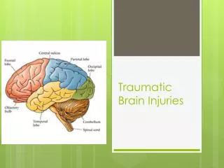
Primary Injury • Acute traumatic intracranial injuries include Primary injurywhich occurs during the initial insult, and results from displacement of the physical structures of the brain.
Secondary Injury • Secondary injury is defined as post-traumatic insults to the brain arising from extracranial sources and intracranial hypertension.
Cerebral Blood Flow • Brain metabolism is dependent on a constant delivery of oxygen and glucose as well as the removal of "waste" products through a constant Cerebral Blood Flow
Cerebral Blood Flow • Cerebral blood flow is equal to the cerebral perfusion pressure (CPP) divided by the cerebrovascular resistance (CVR): CBF = CPP / CVR
Cerebral Perfusion Pressure • Cerebral perfusion pressure (CPP) is defined as the difference between mean arterial and intracranial pressures. • The Brain Trauma Foundation now recommends that the CPP target after severe TBI should lie between 50–70mmHg.
Clinical Correlate • A reasonable estimate of CPP can be made in head injured patients who are not sedated: • Drowsy and confused: (GCS 13-15)ICP=20 mmHg, • Severe brain swelling (GCS <8) ICP=30 mmHg
Clinical Correlate • Thus in a confused, restless and drowsy patient It would be reasonable to estimate his ICP to be 20 mmHg. • A drop in SBP to 80 mmHg drops MAP to 65 mmHg and therefore CPP falls to less than 45 mmHg.
Cerebral Vascular Resistance • CVR is controlled by four major mechanisms: • Pressure autoregulation • Chemical control (by arterial pCO2 and pO2) • Metabolic control (or 'metabolic autoregulation') • Neural control
Pressure Autoregulation • In the normal brain, when the MAP is between 60 and 150 mm Hg, cerebral vessels work to maintain desirable CBF through their ability to constrict and dilate. This is termed “autoregulation.”
cont… • When the MAP is less than 50 mm Hg or greater than 150 mm Hg, the arterioles are unable to autoregulate and blood flow becomes entirely dependent on the blood pressure, a situation defined as pressure-passive flow.
cont.. • This process can only be broken by increasing the blood pressure to raise CPP, inducing the vasoconstriction cascade.
Neuronal Control • Cushing Reflex - is a hypothalamic response to ischemia, usually due to poor perfusion in the brain.
cont… • The ischemia activates the sympathetic nervous system, causing an increase in the heart's output by increasing heart rate and contractility along with peripheral constriction of the blood vessels.
cont… • The increased blood pressure also stimulates the baroreceptors (pressure sensitive receptors) in the carotids, leading to an activation of the parasympathetic nervous system, which slows down the heart rate, causing the bradycardia
Mechanisms of Secondary Brain Injury • Mechanisms that lead to secondary brain injury are: • Hypoxia • Hypotension • Increased intracranial pressure • Hypercarbia • Acidosis
Signs of Herniation • GCS of three to five. • Abnormal posturing - a characteristic positioning of the limbs indicative of severe brain damage. • One or both pupils may be dilated and fail to constrict in response to light. • Vomiting can also occur due to compression of the vomiting center in the medulla oblongata.
Medical Therapy For Increased ICP • The indication for treatment of elevated ICP with hyperosmolar therapy is for short-term treatment while further diagnostic procedures (CT scan of the brain) and interventions (such as treatment of mass lesion found on CT scan) are performed.
Hypercarbia Hypercarbia
Hyperventilation to Reduce ICP • Thus hyperventilation can lead to a mean reduction in intracranial pressure of about 50% within 2-30 minutes. • When PaCO2 is less than 25 mmHg (3.3kPa) there is no further reduction in CBF.
Hyperventilation to Reduce ICP • Acute hypocapnic vasoconstriction will only last for a relatively short time (5 hours) due to a gradual increase in CBF towards control values leading to cerebral hyperaemia (over-perfusion) if the PaCO2 is returned rapidly to normal levels
Contributing Events In The Pathophysiology Of Secondary Brain Injury
Independent Predictors of Poor Outcome • Age • Head CT intracranial diagnosis • Pupillary reactivity • Post-resuscitation GCS • Presence or absence of hypotension.
1. Age • There is an increasing probability of poor outcome with increasing age, in a stepwise manner with a significant increase above 60 years of age.
2. Head CT Intracranial Diagnosis • Initial CT examination demonstrates abnormalities in approximately 90% of patients with severe head injury. • Prognosis in patients with severe head injury with demonstrable pathology on initial CT examination is less favorable than when CT is normal.
2. Head CT Intracranial Diagnosis • Individual CT characteristics found to be particularly relevant in terms of prognosis were: • Compressed or absent basal cisterns measured at the midbrain level. • tSAH • Blood in the basal cisterns • Extensive tSAH
2. Head CT Intracranial Diagnosis • Individual CT characteristics found to be particularly relevant in terms of prognosis were: • Presence and degree of midline shift at the level of the septum pellucidum • Presence and type of intracranial lesions
3. Head CT Intracranial Diagnosis Marshall Classification of Diffuse Brain Injury • Grade 1 = normal CT scan (9.6% mortality) • Grade 2 = Basal cisterns present, shift < 5mm (13.5% mortality) • Grade 3 = Basal cistern compressed/ absent, shift <5mm (34% mortality) • Grade 4 = Shift > 5mm (56.2% mortality)
4. Pupillary Reactivity • The parasympathetic, pupilloconstrictor, light reflex pathway mediated by the third cranial nerve is anatomically adjacent to brainstem areas controlling consciousness.
4. Pupillary Reactivity • Pupillary size (<4mm) and light reflex (>1 mm) are indirect measures of dysfunction to pathways subserving consciousness and, thus, an important clinical parameter in assessing outcome from traumatic coma.
5. Post-Resuscitation GCS • If the initial GCS score is reliably obtained and not tainted by prehospital medications or intubation, approximately 20% of the patients with the worst initial GCS score will survive and 8%-10% will have a functional survival.
6. Presence or Absence of Hypotension • A systolic blood pressure less than 90 mm Hg was found to have a 67% PPV for poor outcome and, when combined with hypoxia, a 79% PPV.
6. Presence or Absence of Hypotension • A single episode of hypotension (SBP <90 mm Hg) is associated with doubling of mortality and increased morbidity when compared to similar patients without hypotension.
Complications • Skull base fracture – CSF leak • Depressed skull fractures – infection risk • Pneumocephalus
Complications • Traumatic subarachnoid haemorrhage • Chronic subdural haematoma • Epilepsy
Complications • Hydrocephalus • Cranial nerve trauma • Concussion • Post-traumatic encephalopathy after repeated injury
References • An Evidence-Based Approach To Severe Traumatic Brain Injury – Emergency Medicine Practice; December 2008 Volume 10, Number 12 • Head Injury - A Multidisciplinary Approach; Edited by Peter C. Whitfield Consultant Neurosurgeon and Honorary Clinical Senior Lecturer South West Neurosurgery Centre Derriford Hospital Plymouth Hospitals NHS Trust Plymouth, UK