Head Injuries
Head Injuries. Dr. S.R. Hulathduwa MBBS , DLM.MD . DMJ(Path) ( Lond .) DMJ( Clin )( Lond .) Dip.Crim . MFFLM(UK ) Senior Lecturer Department of Forensic Medicine University of Sri Jayewardenapura. Regional Injuries. This includes Head injuries Neck injuries Facial injuries

Head Injuries
E N D
Presentation Transcript
Head Injuries Dr. S.R. Hulathduwa MBBS, DLM.MD. DMJ(Path) (Lond.) DMJ(Clin)(Lond.) Dip.Crim. MFFLM(UK) Senior Lecturer Department of Forensic Medicine University of Sri Jayewardenapura
Regional Injuries • This includes • Head injuries • Neck injuries • Facial injuries • Chest injuries • Abdominal injuries • Musculoskeletal injuries
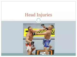
Head Injuries • An important topic in clinical forensic medicine and forensic pathology. • Commonly encountered in traffic accidents, assaults and falls. • Any weapon may be used (cluds, swords, sticks, stones, firearms, bombs..etc.)
Force applied may be • Direct trauma (force directly applied to head) e.g. assaults, accidents, falls • Indirect trauma (transmitted force) e.g. fall off height landing on feet or buttocks, blow to chin, hitting the ground of a motor cyclist in RTA • Acceleration/deceleration e.g. violent shaking(shaken body syndrome) RTA
The force may be • Translational e.g fall • Rotational e.g. blows, shaking, etc..
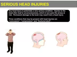
The following anatomical peculiarities modify the nature and the extent of head injuries. - hair, scalp, skull, meninges, brain and base of skull HOW? e.g. scalp bruises are better felt than seen. -Bleed profusely even PM. -Easy spread of infection. -Sinus thrombosis.
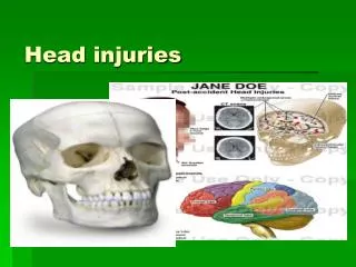
Types of head injuries • Hair – cuts, crushed bulbs and burns. • Scalp – abrasions, lacerations, bruises incised wounds, stabs, burns • Sub scalparhaematoma • Pericranial haemorrhages. • Skull-fractures, suture diasthesis, cuts, deficiencies. • Extra duralheamorrhages(EDH) • Dural tears • Sub dural haemorrhages (SDH) - acute, sub acute, chronic
Pia-arachnoid tears. • Sub arachnoid heamorrages (SAH) • Brain-contusions, lacerations, cuts, stabs, intra cerebral haemorrhages, intra ventricular haemorrhages. • Brain stem – primary and secondary brain stem heamorrhages. • Damage to cerebral blood vessels. • Raised intracranial pressure
Scalp injuries • Layers of scalp S- C- A- L- P-
Abrasions • Are rare due to presence of head hair. • May also be unidentified/ missed due to presence of head hair. • May indicate severe friction unless the person is bald. eg. RTA, fall off height. etc..
Bruises / Contusions • May be hidden by hair. • Occurs between the aponeurosis and skin. • Better felt than seen • Felt as a boggy mass, swelling or a bump, specially when associated with a sub-scalparhaematoma. • Well seen in autopsy when the scalp is reflected.
May give the false sense of a fracture of the underlying skull on external palpation. • May gravitate to appear at a distant site (eg. Black eye) • May become confluent with time so that it will not accurately indicate the actual number of blows dealt.
Lacerations • Usually spilt lacerations due to compression of scalp between a blunt object and underlying skull: thus resembling incised wounds. How to differentiate? • Depending on the offending object/ weapon, there may be other types of lacerations too (crush / perforating / blast lacerations) • May sometimes give a clue to the shape of the offending weapon. eg. Stellate – blunt rods /falling backwards.
Incised wounds • Usually slashed cuts. Stabs -usually on an area where the skull is thin. E.g. temporal bone. -also can penetrate the orbit.
Burns • Could be due to flame / high tension electrocution / corrosives. • Flame burns may cause singeing of hair, which is of value in, a)Deciding the approximate range of fire arm discharge. b) Deciding the proximity of the victim to the explosion. c) Differentiating dry heat from scalds (moist burns) and corrosive burns
Fire arm injuries -perforating and/or blast lacerations. Blast injuries -blast and/or perforating lacerations.
Skull fractures • A fall through one meter (or even lesser) can cause a skull fracture in an adult. • Usually a substantial force in needed to cause a skull fracture. • May involve one or both tables of the skull. • Common on the vault than the base of the skull.
Types of skull fractures • Linear / fissured • Depressed • Comminuted • Depressed comminuted • Guttered • Elevated • Growing • Pond • Ring • Compression • Hinge • Contre-coup
Linear/fissured # -usually falls. -also by blows with blunt weapons on an unsupported head. -may represent the forward and downward components of the force applied, thus indicating the direction of the blow/position of the victim. (pointer #)
Depressed # • When the inner table is disrupted and driven in. • Usually due to blows with a heavy weapon with a relatively small striking surface. E.g. head of a hammer • Common with supported head. • May usually indicate the shape of the striking surface, thus known as “signature fracture”
Damage to meninges and brain is common. • If the force is severe enough, can end up with a deficiency of the skull. - Can be a focus of post traumatic epilepsy at a later time.
Comminuted # • When the skull is broken in to more than two fragments. • May take the shape of a “cob-web” )cob-web/mosaic #) e.g. RTA, falls, blunt heavy weapon with a large striking surface. • Depressed comminuted #
Guttered # • An elongated depressed # e.g. long, heavy, thin, blunt weapon. • Elevated # - When a cutting/stabbing weapon is withdrawn from the skull, the fractured fragments may be elevated.
Pond # • In infants the skull may bend / indent inwards without “ cracking” • Growing # - If the soft tissue/meninges are caught in between the two edged of a fracture, in an infant, the # will widen with time delaying healing. Brain tissue may even herniate through the defect with time.
Ring # - Around the foramen magnum. • Usually due to transmitted force along the spine (fall off height landing on feet/buttock) • Rarely when landing on the top of head. • Compression # • Due to lateral compression of head. • The fracture line may encircle the head.
Hinge # • Fracture line runs across the middle cranial fossa and pitutaryfossa dividing the base of the skull in to two. • Seen when motorcyclists land on the road chin first (motorcyclist’s #) • Also in boxers.
Contre-coup # • Orbital plate of the frontal bones (orbital roofs) and ethmodial plate may be fractured due to occipital impact. Eg. Fall on the back of the head. • Any of the above can be compound internally or externally.
Factors modifying the nature of a skull fracture • Nature of the weapon/agent (blunt, sharp, cutting, stabbing, firearm, sharpnel) • Striking surface • Pliability of the weapon • Force used
Thickness of the skull • Area of the head involved (e.g temporal Vs occipital) • Whether the head was supported or free to move. • Other factors related to the victim (eg. Age, bone diseases, etc..)
Forensic information that may be gathered from skull fractures. • Nature of the weapon/agent. • Force used(a crude approximation only). • Whether the head was supported or free to move. • Direction of the blow(by studying the pointer #) • Possible external injuries • Possible internal injuries • Volitional activities.
Period of survival • COD • Evidence of infection/healing • Is it fall or a direct blow? HOW? e.g. depressed # on the vertex are usually due to a direct blow while a linear # in occipital area. Extending in to base of skull is more suggestive of a fall backwards.
Intra cranial haemorrhages This includes, • Extra dural haemorrhages (EDH) • Sub dural haemorrhages (SDH) (acute, sub acute, chronic) • Sub arachnoid haemorrhages (SAH) • Intra cerebral haemorrhages • Ventricular haemorrhages
Cerebral injuries The following can be included as cerebral injuries, • Cerebral concussion • Diffuse axonal injuries (DAI) • Diffuse vascular injuries • Coup injuries • Contre-coup injuries • Intra cerebral haemorrhages • Brain stem haemorrhages • Ischaemic brain damage (hypoxic ischaemic encephalopathy)
Extra Dural Haemorrhage(EDH) • Collection of (clotted) blood in the potential space between the inner table of the skull and the dura. • Commonest site is the temporal area as the skull is thin and the meningeal vessels run in this area. • Parietal is the second commonest. • Can rarely be infra tentorial in the posterior cranial fossa
Symptoms occur rapidly in temporal and parietal EDH, enabling early diagnosis. • Can present in 3 ways. 1.) classical presentation (yet rare) immediate loss of consciousness Spontaneous recovery with lucid interval Gradual deterioration of level of consciousness until death
2).No LOC initially gradually deteriorates until death 3). Unconscious from the beginning Dies without regaining consciousness • Prognosis is poor in frontal EDH as onset of symptoms is delayed. • EDH in posterior cranial fossa can cause brainstem compression (infra-tentorial EDH)
EDH is considered to be always traumatic in origin; 85% are associated with skull # • Source of bleeding could be either arterial (middle meningeal) or venous (diploic veins, superior sagittal sinus and other venous sinuses) • Onset is rapid arterial bleeds and in most cases the bleed is arterial. • In an adult, approximately 100-150 ml is considered to be fatal if not removed.
After established cerebral oedema due to increased intracranial pressure, EDH will have a poor prognosis, though successfully evacuated. • Usually considered as FIOCN. • Rare in extremes of age: why? • Bilateral EDH can occur with compression # though this is very rare. • “heat haematoma” is a post mortem artifact due to burns, which will resemble an ante-mortem EDH
When the skull of the dead body is subjected to intense heat, blood will seep from the diploe and collect above the dura. • Usually seen underneath an area of extensive burns: may be bilateral and chocolate brown in colour (or even cherry pink due to carboxy-haemoglobin), Honey-comb/cavernous in appearance: • But problem may occur when associated with other heat artifacts such as ruptures as heat ruptures and heat fractures.
Subdural haemorrhage (SDH) • A collection of blood between dura and piaarachnoid. • Commoner than EDH. • Less commonly associated with skull fractures when compared with EDH • May occur over convexities of the brain as well as in all three cranial fossa. • Almost always traumatic. Rarely non traumatic due to rupture of an aneurysm in the posterior communicating artery; How?
Also burst lobe following a spontaneous intra cerebral haemorrhage, is also non traumatic. • Bleed is mainly venous (bridging veins, dural sinuses, extension of an intra-cerebral haemorrhage as a burst lobe). • Clinically can be, -acute -sub acute -chronic
Usually does not occur as an isolated injury, thus category of hurt is not clear cut. • Chronic SDH (an organized sub dural blood clot) can be an incidental finding in the elderly at the autopsy. • Clinical effects of a chronic SDH can be misinterpreted as those due to neuro-psychiatric disorders.
A chronic SDH will be seen as a brownish, cystic, gelatinous mass containing fluid of various colours (red/brown/yellow), adherent to dura and covered with a neo membrane. • This membrane usually grows from the dural surface. The degradation of blood pigments gives the different colours. • Munro has devised a dating system (ageing of the chronic SDH) by histological examination of the neo membrane.
SDH is common in extremes of age. E.g. infants – shaken baby syndrome -elderly – due to shrunken brain with widened sub dural space. • Also in, -chronic alcoholics punch drunk syndrome -boxers
Category of hurt is variable -may be FIOCN • In a criminal trial (eg. Assualt case) it may be important to prove/disprove the causative relationship of the assualtive event and the SDH, which has resulted in death. This may be attemped by histoligical ageing of the neo membrane.
Sub archonoid haemorrhage (SAH) • Collection of blood between pia and arachonoid. • Can be spontaneous or traumatic. • Spontaneous; • Rupture of berry aneurysm (read path notes) • Extension of a spontaneous intacerebral or an intraventricular haemorrhage in to sub arachonoid space as a burst lobe or through the normal path of CSF flow.
Traumatic 1. Primary traumatic due to diffuse rotational trauma to brain, bridging vessels are ruptured. The SAH is diffuse. There may be other associated injuries such as SDH, contre-coup injuries. 2. Secondary traumatic due to localized trauma to brain (eg. A cerebral contusion or a laceration). There will be localized SAH in that area.
3. Extension of a traumatic intra-cerebral or/and intra ventricular haemorrhage in to sub arachonoid space. 4. Basal SAH can occur due to lateral neck trauma resulting in the rupture of vertebral vessels (how to demonstrate a vertebral artery damage in the autopsy?) 5. Sudden fluctuation of the pressure in the neck arteries without actual trauma to vessels in the neck. e.g. sudden compression of the neck in manual strangulation.
Categorization of hurt is difficult when associated with other injuries. May depend on the individual case. • It may be an important (yet very difficult) issue to link a rupture of berry aneurysm causing a traumatic SAH following an assaultive event. Read clinical presentation.
Concussion • An increasingly important topic in clinical forensic medicine and sports medicine. • Different authors classify concussion in to different grades. Eg. CANTU classification Colarado classification AAN classification • Grade I,II,III etc.