Head injuries
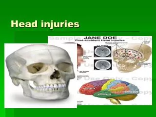
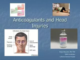
Head injuries. Definition. Head injury is an injury to the scalp, skull, or brain. The most important consequence of head injery is traumatic brain injury.

Head injuries
E N D
Presentation Transcript
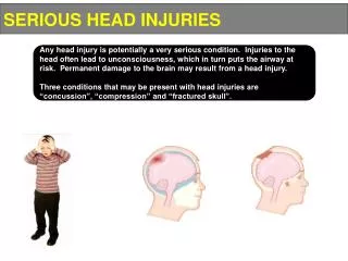
Definition • Head injury is an injury to the scalp, skull, or brain. • The most important consequence of head injeryis traumatic brain injury. • Head injury may occur either as a closed head injury, such as the head hitting a car or as a penetrating head injury, as when a bullet pierces the skull. Both may cause damage that ranges from mild to severe . • Very severe injury can be fatal because of profound brain damage.
Possible injuries to Brain • Cerebral Contusion Is the commonest form of traumatic intra-axial injury. • Contusions occur at the inferior and polar surfaces of the frontal and temporal lobes. • Cerebral contusions are also produced secondary to depressed skull fractures and are associated with other intracranial injuries
Clinical features • Brain injury may be generalized with widespread brain damage ( In this case an increase in the vascular permeability will occur resulting in cerebral edema • decreased respiratory exchange in severely injured patients also leads to anoxia resulting in cerebral vasodilatation which lead to cerebral swelling • Usually associated with a brief loss of consciousness. • The elderly patients, alcoholics and patients taking anticoagulants are at increased risk of hemorrhage
Radiological features • Non-contrast computed tomography (CT) useful in the early posttraumatic period. • Contusions are seen as multiple focal areas of low or mixed attenuation intermixed with tiny areas of increased density representing hemorrhage. • True extent becomes apparent over time with progression of cell necrosis and edema. • Magnetic resonance imaging (MRI) is the best modality for demonstration of edema and contusion distribution. Cerebral contusions in both frontal lobes (arrows). The adjacent low density represents local edema.
Skull fractures • Skull fractures are more serious than other bones fractures if the brain is injured • These fractures can be classified into :- Closedif the overlying skin is intact OR Compoundif the overlying skin is affected Linearif there is a single fracture line Stellateif there is several fracture lines radiating from a central point Depressed or not depressed according to the movement of the fracture edges below the level of surrounding bone
Infra-orbital fracture. (a) ‘Teardrop’ sign. (b) Coronal CT demonstrating the same. a)) (b)
Simple Skull fractures • This may be linear- stellate or comminuted but not depressed • if they cross the major vascular channels epidural and subdural haematoma may occurs • the patient with this type of fractures should be kept under close observation until certain that no bleeding is occurring )Simple vault fracture (arrowheads
Depressed Skull fractures • Depressed fractures affects the underlying brain cortex which may cause neurologic effects • Some time called ping-pong fractures • A tangential view may use to show the degree of depression if CT unavailable
Skull Base fractures • Are fractures through the dense inner structures of the temporal bone • the major signs includes rhinorrhea , otorrhea • this type of fractures is very difficult to demonstrate according to the complex anatomy through this area. • If bleeding occurs plain radiographs may revel air-fluid level in the sphenoid sinus ( lateral view) • the use of CT scan is more effective to demonstrate this type of fractures skull base fracture
Gunshot wounds • Gunshots can be visualized by plain film images which used to localize the bullets in gunshot victim ( the bullet is easily localized because of lead contents Scalp injuries • Scalp is an extremely vascular structure , and injury may cause serious hemorrhage • All scalp wounds should be closed as soon as possible unless they overlying a depressed fracture or penetrating wound of the skull which need special care in the operating room • The common Scalp injury is laceration
investigations • Skull films are not indicated routinely for the following indications: • Headache • Possible pituitary problems - ( CT/MRI preferred) • Possible space occupying lesion • Epilepsy • Dementia or memory loss • Middle or inner ear problems • Temporal mandibularjoint dysfunction - will not show disc abnormality which is the most common cause of dysfunction.
Subarachnoid haemorrhage Intracerebral hematoma
Large hypo dense area Brain infarct
Epiduralhematoma Subdural hematoma