UNIT III – CELL STRUCTURE & FUNCTION
Discover the history of microscopes, cell theory, and the types of microscopes used to study cells. Learn about prokaryotic and eukaryotic cells, their structures, functions, and the components of eukaryotic cells. Explore the endomembrane system, the roles of organelles, and cell boundaries such as the cell wall and extracellular matrix.

UNIT III – CELL STRUCTURE & FUNCTION
E N D
Presentation Transcript
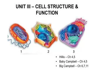
UNIT III – CELL STRUCTURE & FUNCTION • Hillis – Ch 4,5 • Baby Campbell – Ch 4,5 • Big Campbell – Ch 6,7,11
I. DISCOVERY OF CELLS • History of Microscopes • Anton van Leeuwenhoek • Robert Hooke • Cell Theory • All living things are made of cells. • Cells are the smallest working unit. • All cells come from pre-existing cells through cell division.
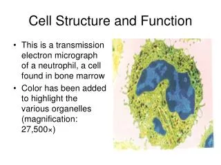
I. DISCOVERY OF CELLS, cont. • Types of Microscopes • Compound Light Microscope • Magnification • Resolution • Advances in light microscopy include • Confocal • Fluorescent • Phase Contrast • Super-resolution • Electron Microscope • Scanning Electron Microscope (SEM) • Transmission Electron Microscope (TEM)
I. DISCOVERY OF CELLS, cont. • Cell Size • Metabolic needs impose both upper & lower limits on cell size • How small? • Must have enough space for DNA, enzymes • Mycoplasma sp. - < 1 μm • How large? • Surface Area to Volume Ratio • Adaptations
II. CELL TYPES • Prokaryotic Cells • Typically smaller than euks • Bacteria • Kingdom • Kingdom • No true nucleus – DNA found as a single chromosome in region called nucleoid
II. CELL TYPES, cont • Prokaryotic Cells
II. CELL TYPES, cont • Eukaryotic Cells • Larger, more complex • Contain true nucleus, membrane-bound organelles suspended in cytosol • Composed of • Nucleus • Ribosomes • Endomembrane System • ER • Golgi Apparatus • Lysosomes • Vacuoles • Mitochondria/Chloroplasts • Peroxisomes • Cytoskeleton
III. EUKARYOTIC CELL STRUCTURES _______________ Cell _____________ Cell
III. EUKARYOTIC CELL STRUCTURES, cont • Control center of eukaryotic cell • Double membrane that protects nucleus; continuous with ER • Contains pores • Site of ribosome production • DNA wrapped in protein
III. EUKARYOTIC CELL STRUCTURES, cont • Suspended in cytosol or found on rough ER • Site of protein production in a cell
III. EUKARYOTIC CELL STRUCTURES, cont • Endomembrane System • Endoplasmic Reticulum • Interconnected network continuous with nuclear envelope • Rough ER • Smooth ER
III. EUKARYOTIC CELL STRUCTURES, contEndomembrane System, cont • “Cell postmaster” • Receives transport vesicles from ER; modifies, stores, and ships products • Receiving side is known as the cisface; shipping side is known as the trans face
III. EUKARYOTIC CELL STRUCTURES, contEndomembrane System, cont • Sacs containing hydrolytic enzymes • Used for recycling cellular materials, destroying pathogens
III. EUKARYOTIC CELL STRUCTURES, contEndomembrane System, cont • Storage sac • Plants typically have large, central vacuole surrounded by membrane called tonoplast. Absorbs water and helps plant cell to grow larger • Some protists have contractile vacuole to pump out excess water
III. EUKARYOTIC CELL STRUCTURES, cont • Site of oxidative respiration • Contain own DNA, ribosomes • Found in virtually all euk cells • Enclosed by 2 membranes; inner membrane has folds called cristae to increase surface area
III. EUKARYOTIC CELL STRUCTURES, cont • Type of plastid that carries out photosynthesis by converting solar energy to chemical energy (glucose) • Contain membranous system of flattened sacs called thylakoids – stack is called a granum • Fluid surrounding thylakoids is called stroma • Contains DNA, ribosomes
III. EUKARYOTIC CELL STRUCTURES, cont • Membrane-bound compartments that use O2 to carry out metabolism • H2O2 is produced; broken down by _________________
III. EUKARYOTIC CELL STRUCTURES, cont • Provides structural support to cell • Allows for movement • Attachment site for organelles, enzymes • More extensive in animal cells • Composed of three types of proteins • Actin • More fixed • Keratin
IV. CELL BOUNDARIES • Cell Wall • Found in • Rigid structure; protects, maintains shape of cells • Prevents excess water uptake • Plant cell wall • Cellulose • Pectin - Sticky polysaccharide found between cell walls of adjacent cells • Plasmodesmata - Perforations between adjacent cell walls that allow for movement of materials from one cell to another
IV. CELL BOUNDARIES, cont • Extracellular Matrix of Animal Cells • Holds cells together, protects & supports cells • Allows for communication between cells • Composed primarily of glycoproteins – proteins with covalently-bonded carbohydrate chains attached • Must abundant glycoprotein in most animals is collagen
IV. CELL BOUNDARIES, cont • Intracellular Junctions in Animal Cells • Tight Junctions – Press membranes together very tightly; prevents leakage of fluid • Desmosomes (Anchoring Junctions) – Fasten cells together in sheets • Gap Junctions – Allow for movement of cytoplasm from one cell to another; important in communication between cells
IV. CELL BOUNDARIES, cont • Cell (Plasma) Membrane • Selectively-permeable barrier found in all cells • Composed primarily of phospholipid bilayer • Fluid Mosaic Model • “Fluid” – Not a rigid structure. Organization due to high concentration of water inside & outside cell
IV. CELL BOUNDARIES, cont • Organization of Plasma Membrane
IV. CELL BOUNDARIES, cont • Fluidity of Plasma Membrane
IV. CELL BOUNDARIES, cont • Cell Membrane, cont • Proteins - “Mosaic” – Assortment of different proteins embedded in bilayer; determine most of membrane’s specific functions. Act as channels, pumps, enzymes in metabolism, binding sites, etc • Integral Proteins – Embedded in phospholipid layer • Peripheral Proteins – Bound to surface of membrane
IV. CELL BOUNDARIES, cont • Cell Membrane, cont • Carbohydrates • “ID tags” that identify cell. • Enable cells to recognize each other and foreign cells. • May be bonded to lipids (glycolipids) or proteins (glycoproteins)
V. CELL TRANSPORT, cont • Passive Transport – Movement of materials from high to low concentration. No energy output required. • Diffusion • Random movement of a substance across membrane down concentration gradient • No net movement once equilibrium is reached
V. CELL TRANSPORT, cont • Passive Transport, cont • Facilitated Diffusion • Passive transport of molecules across cell membrane with the help of transport proteins • Utilized by large molecules, charged particles, polar molecules • Water
V. CELL TRANSPORT, cont • Passive Transport, cont • Osmosis – Diffusion of water across a membrane. Tonicity refers to tendency of cell to gain or lose water. If the solution is • Isotonic relativeto the cell – Solute concentration is same on both sides of membrane. No net movement of water. • Hypertonic relative to the cell – Concentration of solute is greater outside cell → water moves out of cell until equilibrium is reached. Cell may shrivel. • Hypotonic relativeto the cell – Concentration of solute is lower outside cell → water moves into cell until equilibrium is reached. Cell may swell to bursting point.
V. CELL TRANSPORT, cont • Passive Transport / Osmosis, cont • Water Potential • Used to predict the passive movement of water • Designated as Ψ • Water always moves from an area of higher water potential → lower water potential • ΨS = • ΨP =
V. CELL TRANSPORT, cont • Passive Transport/Osmosis, cont • Osmoregulation • Cells must have mechanism to prevent excess loss, uptake of water • Cell wall, contractile vacuole • Plasmolysis – Seen in plants; excessive water loss causes cell membrane to pull away from cell wall
V. CELL TRANSPORT, cont • Active Transport • Movement of materials against concentration gradient. Requires energy output by cell • Carrier Proteins – Na+ / K+ Pump
V. CELL TRANSPORT, cont • Active Transport, cont • Proton Pump
V. CELL TRANSPORT, cont • Active Transport, cont • Exocytosis • Secretion of biomolecules by fusion of vesicles with cell membrane. Biomolecules “spit out”. • Hormones, neurotransmitters, etc
V. CELL TRANSPORT, cont • Active Transport, cont • Endocytosis – “Sucking In”. Cell membrane surrounds, engulfs particle or biomolecule, pinches in to form vesicle. • Phagocytosis – “Sucking in” food particles • Pinocytosis – “Sucking in” fluid droplets • Receptor-mediated Endocytosis – Very specific
VI. CELL SIGNALING • Autocrine Signaling
VI. CELL SIGNALING, cont • Coordinates cell activities, development • Typically involves 3 steps: • Reception – Target cell’s detection of signal molecule due to binding of signal molecule to receptor protein in cell membrane • Transduction – Binding of signaling molecule changes receptor protein; triggers a sequence of events within cell • Response – Results in specific cellular response; for example, activation of genes, enzyme catalysis, etc.
VI. CELL SIGNALING, cont • Reception • Typically involves G Proteins
VI. CELL SIGNALING, cont • Transduction • Typically multi-step pathway • Relay molecules are usually protein kinases
VI. CELL SIGNALING, cont • Transduction • Non-protein molecule known as cAMP is often second messenger
VI. CELL SIGNALING, cont • Response • Nuclear • May “turn on” or “turn off” genes • Cytoplasmic • May regulate enzyme activity • Apoptosis • Controlled cell suicide
VI. CELL SIGNALING, cont • Regulation