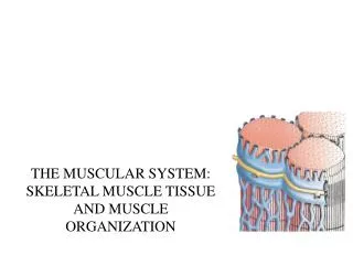
Understanding Muscle Properties: Contractibility, Excitability, Extensibility, and Elasticity
Muscle tissue exhibits four fundamental properties: contractibility, excitability, extensibility, and elasticity. Contractibility allows muscle cells to shorten and generate force, while excitability enables them to respond to stimuli, such as nerve impulses and hormones. Extensibility refers to the ability to stretch without losing function, and elasticity allows muscles to return to their original shape after contraction. There are three main types of muscles: skeletal, cardiac, and smooth, each with distinct structures and functions essential for body movement and organ function.

Understanding Muscle Properties: Contractibility, Excitability, Extensibility, and Elasticity
E N D
Presentation Transcript
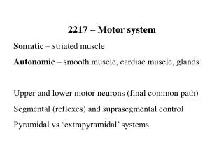
Muscle System Ch 9
Muscle Properties • 4 basic properties • Contractability • Excitability • Extensibility • Elasticity
Contractability • Cells capable of decreasing along a longitudinal axis • Shorten & thicken • Produce force • Pull or create tension
Excitability • Ability to respond to external stimulation • Stimulation initiated by • Hormonal cues • nerves • Motor nerves
Extensibility • Ability to “stretch” without loss of function
Elasticity • Ability to regain original shape following contraction
Muscle Types • Skeletal • Cardiac • Smooth
Skeletal • Aka muscle fiber • Primary muscle type • 700+ • Voluntary • Only consciously controlled tissue • Move & stabilize the skeleton • Striated • Contractile proteins produce movement (contraction), striated in appearance • Large, multinucleate cells • Long thin cells= myofibers • Some regeneration
Skeletal Muscle Function • By mean of contractions..
Skeletal Muscle Functions • Produce skeletal movement • Maintain posture & position • Support soft tissue (pelvic floor & abdominal wall) • Regulate entering & exiting of material • Digestive & urinary tract • Thermogenic • Produce body heat
Motor Control • Controlled by higher brain regions (Cerebrum) • Allow for conscious control of muscles
Smooth • Aka visceral • Non-striated aka smooth • Involuntary • Not consciously controlled • Small spindle shaped cells • Microfibers with random arrangement • Uninucleate • Regenerative • Functions in transporting fluids & solids through the body • Ex digestive system, urinary structures, blood vessels, glands, reproductive tract
Cardiac • Involuntary striated • Self stimulating • Only in heart • Small, uninucleate, interconnected, branched cells • Intercalated discs • Visible cellular connections • Gap junctions • Desmosomes • Functions to push blood through blood vessels • No regeneration
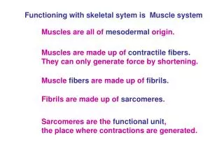
Skeletal Muscle Anatomy • Specialized cells- Myofibers • Long slender fiber like cells • Mature cells multinucleate • NOT capable of mitotic division • Cellular development • Fusion of many embryonic stem cells form long multinucleate cells • myoblasts
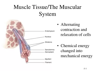
Myofiber Structure • Sarcolemma • Myofiber cell membrane • Sarcoplasm • Myofiber cytoplasm • Made up of bundles of myofibrils • Made up of micrfilaments aka myofilaments • Satallite cells • Muscle stem cells • Resident myoblasts in adult tissue • Tissue repair • Fascicle • Connective tissue holds together for organization • Contains bundles of myofibers
Myofibers are made of bundles of Myofibrils Aka Fascicle
Muscle Connective Tissue • Connective tissue surrounds, supports, & attaches muscle • 3 layers of connective tissue • Endomysium • Perimysium • Epimysium
Endomysium • “Within” • CT surrounding & binding together individual myofibers • Delicate network of reticular fibers • Holds myofibers together • Supports blood vessels • House satellite cells
Perimysium • “around” • CT surrounding & binding groups of myofibers • Fascicles • Stringiness of meat • Collagen & elastic fibers • Houses blood vessels & nerves
Epimysium • “above” • Dense irregular CT • Surrounds & binds fascicles together • Holds together Individual muscles into discrete units • Ex biceps, triceps, deltoid
Epimysium holds together Individual muscles into discrete units
Tendons & Aponeurosis • CT attaching • Muscle to bone • Muscle into CT of another muscle • Combination of CT fibers from all levels of muscle organization • Endomysium • Perimysium • Epimysium • CT fibers are continuous w/ periosteum & osseous matrix= strong muscle attachments • Bones more likely to break before tendon tears away from bone • All CTs merge to form attachments • Tendon- strong cord or rope • Aponeurosis- flattened sheet