Muscle system
Muscle system. Characteristics of the muscle system. Excitable : muscles will respond to stimuli (neural stimuli) Contractile : muscles can contract (shorten) Will do so when excited Extensible : muscles can be stretched

Muscle system
E N D
Presentation Transcript
Characteristics of the muscle system • Excitable: muscles will respond to stimuli (neural stimuli) • Contractile: muscles can contract (shorten) • Will do so when excited • Extensible: muscles can be stretched • Elastic: muscles, when relaxed, will recoil or return to their original position • Think…rubber that will shrink on command
Functions of the muscle system • Act to control of move the skeleton • Mobility, “actions”, communication • Plays a major role in body heat production • 85% of your body heat is generated by skeletal muscle • All your muscle tissue amounts to the second largest user of glucose and oxygen in your body • Blood flow • Muscle contractions help to push blood to the extremities (appendages)
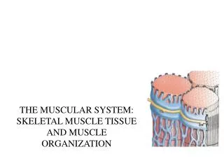
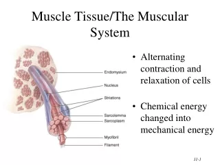
Muscle cells • Also called muscle fibers or myocytes • “Myo” = muscle • Plasma membrane is unique • Sarcolemma (“Flesh lining”) • Plays a role in excitability (similar to a neuron) • Cytoskeleton is a major constituent of skeletal muscle cells • Actin (microfilaments) arranged into bundles • Normally “covered” by “control” proteins that limit contraction
Muscle cells Actin = round balls (in reality, it looks like a bunch of arrowheads stuck in a line) Tropomyosin = “block” or “plug” protein that covers the actin filament Troponin = regulator for tropomyosin
Muscle cells • Interspersed between the actin bundles are thicker bundles of motor proteins = myosin • Looks like a golf club or music note • “head” will grab onto actin and pull itself along the actin filament
Muscle cells • Thick (myosin) and thin (actin) filaments are arranged into a bundle = “sarcomere”
Muscle cells • Sarcomere = the bundles of actin & myosin between “Z-discs” • Z-disc is what holds the actin microfilaments together • Z-disc is also what is attached to the sarcolemma (muscle cell plasma membrane) • Z-disc attached to sarcolemma using “dystrophin” protein • There are 1000’s of sarcomere structures along the length of a muscle cell • Why skeletal muscle also called “striated muscle”…striations = part of the sarcomere
Muscle cells • Dystrophin = muscular dystrophy • 2 kinds of muscular dystrophy • Dystrophy = muscular weakness • Most common forms = genetic disorders • Duchenne muscular dystrophy • Nucleotide sequence for dystrophin often has an extra or missing nucleotide • “out of reading frame”….remember the triplet codon system on DNA? • Becker muscular dystrophy • Protein “primary sequence” off by 1 or 2 amino acids • Protein is made with correct number of amino acids, but 1-2 have been “substituted” due to mutation of change in the DNA sequence
Muscle cells • Muscular dystrophy • Without the correct “connection” between the contracting cytoskeleton and the outer plasma membrane (sarcolemma), the cell will rip itself from the inside out • Cytoskeleton contracts, but isn’t attached to sarcolemma • Repeated back & forth motion within the cell leads to tears in the cell etc.
Muscle cells • Wrapped around the “sarcomere” (actin & myosin bundles) is a specialized form of endoplasmic reticulum • “sarcoplasmic reticulum”: holds calcium • Calcium is the key to contraction • Ca2 = 2+ charge or very strong “ionic” nature • Doesn’t take very much to change the electrical “gradient” • When stimulated, sarcoplasmic reticulum releases calcium into the sarcomere • ONLY for a fraction of a second (a pulse of calcium) • THEN, it quickly re-absorbs the calcium
Muscle cells When you release calcium from the sarcoplasmic reticulum, the calcium will then bind to troponin, and move the tropomyosinOFF of the actin microfilament. This will then permit the myosin head on the THICK filament to interact with the actin microfilament.
Muscle contraction • Muscle cells will ONLY contract when told to do so by a NEURON • Have to be conscious for a muscle to contract • Without neural connection or stimulation, muscle will remain “dormant” • Sever the neural connection (injury etc.), muscle will cease to “function” or contract • “denervation atrophy” if you don’t use it, you’ll eventually lose it
Muscle contraction • Neuron that controls a muscle cell = somatic motor neuron • 1 neuron can control a number of muscle cells • Stronger muscles (gastrocnemius & soleus of the calf) = 1 neuron controls 1000’s of muscle cells • “control” muscles (eye, hands) have lower ratio: 1 neuron controls 3-6 muscle cells
Muscle contraction • When you want to contract a muscle, you “think” if first • Conscious thought triggers somatic motor neurons in brain to send stimulatory impulses to the target muscle group (electrical impulse) • At the muscle cell, the final “interface” = motor end plate
Muscle contraction • At the motor end plate, the neuron does not physically “touch” the muscle cell • Neuron releases acetylcholine (chemical neurotransmitter…made from amino acid choline) • Acetylcholine then triggers an electrical change in the sarcolemma • Neuron releases “pulses” of acetylcholine • Doesn’t drop a whole ton of it at once
The motor neuron will release pulses of stimulatory acetylcholine. Therefore, muscle cells “contract” or respond to the neural pulses by “twitching” or “transiently contracting” rather than a constant action.
Muscle contraction • After twitch (if the muscle cell only receives 1 stimulatory pulse), the sarcolemma will “recoil” back to it’s original length • Recall the elastic fibers that hold the actin microfilament bundles together on the “Z-disc”
Muscle contraction • If a muscle cell only “twitches” and the motor neuron only releases “pulses” of acetylcholine…how do you contract your muscle so much? • Deliver more pulses • But, neurons have only a limited number of acetylcholine pulses they can deliver • Takes a great deal of energy to make and release acetylcholine • If you fire 1 neuron fast enough, it will exhaust itself (work itself to DEATH)
Muscle contraction • Remember this slide: • Each muscle in your body is made of 1000’s or more muscle cells • Each muscle is therefore “fed” by 100-1000’s of neurons • ALL the neurons that feed a muscle NEVER fire at the same time (you’d break a bone if that happened) • You fire “groups” of neurons • You also fire groups of neurons in succession
Muscle contraction • Firing groups of neurons helps to spread the metabolic load throughout a number of neurons and a number of muscle cells that make up a muscle • For a given contraction (ie. lifting a book), only about 20-70% of the muscle cells are involved • The neurons trigger muscle cells to contract in succession • Like a flip comic book…the faster you flip the pages, the more fluid the motion
You normally have a number of somatic motor neurons that control a number of muscle cells. These neurons trigger contractions in succession. Since all the muscle cells are connected to the same tendon, you can adjust the strength of the overall muscle contraction by adjusting how many muscle cells contract at any one time. Note that you never have EVERY muscle cell contracting at the same time.
Muscle contraction • Spreading the “metabolic load” • Each myosin head needs 1 ATP molecule in order to attach to the actin microfilament and “move” to the next • For each sarcomere, there are 1000’s of myosin heads • 1000’s of ATP molecules per sarcomere… • 1 muscle cell can have 1000’s of sarcomeres!!!! • Each neuron needs a great deal of ATP to perform the exocytosis to release acetylcholine • The sarcoplasmic reticulum needs a great deal of ATP to “vacuum” up the calcium released • This is why you never trigger EVERY muscle cell in a muscle to contract at once (they would all wear out at the same time)
There is a limit to how much activity your muscles can carry out About 6 seconds of “burst” energy is stored as ATP About 10 seconds worth can be “recycled” using the creatine phosphate system Muscle cells then revert to anaerobic fermentation to produce 2 ATP’s from 1 glucose (they have about 30-40 seconds worth of glucose stored for this) After this, (hopefully), your heart has started to pump more blood (with oxygen and glucose) to your muscles to permit them to undergo aerobic respiration
Muscle metabolism • Roughly 6 seconds of “stored ATP” • Once used up, have to make more • For about 4 seconds after all the ATP is used up, your muscle cells can “recycle” ADP into ATP • Uses Creatine phosphate system • Creatine phosphate has PHOSPHATE that can be quickly attached to ADP • Only have about 4-5 seconds worth of creatine phosphate
Muscle metabolism • After creatine phosphate system is exhausted, muscle cell relies on anaerobic fermentation (simply hydrolysing glucose into 2 pyruvate halves) • Muscle cells store glucose • About 30-40 seconds of glucose for anaerobic fermentation • Unlike liver cells, muscle cells store glucose as glycogen, but DO NOT release it into the blood • Ideally, some time during anaerobic fermentation, your heart rate will increase enough to deliver oxygen to your muscle cells to permit aerobic respiration
Muscle “training” • You can “train” your muscles to increase their strength & endurance • Endurance training = increasing the amount of glucose (glycogen) stored in each muscle cell • Increasing bloodflow to the muscles • Increasing red blood cell count (increased oxygen in blood) • Strength training = increasing the creatine phosphate (ATP recycling mechanism) supply • Building muscle mass is largely through “micro-tears” in the muscle cells that heal
Muscle actions • 1 muscle rarely acts alone • In order to move your skeleton (“articulate” a joint), many muscles must combine their efforts
Types of muscle contractions Knowing that twitches are “added” or “wave summated” in order to elicit a contraction, we now want to know what kind of contractions we can elicit Isometric contraction: contraction without a change in muscle length When you hold in one place…it takes muscles to hold your book in front of you, even if none of the muscles in your arm are changing length Isotonic contraction: contraction resulting in a change in muscle length, but no alteration in muscle tension When you increase the internal tension in the muscle to overcome the resistance
Types of muscle contractions Isotonic contraction: 2 forms of isotonic contractions Concentric contraction: muscle shortens, tension remains the same When you lift something with your arm, you concentric contract your biceps brachii Eccentric contraction: muscle lengthens, tension remains the same When you gently place something down with your arm, you eccentrically contract your biceps brachii You have more strength with this form of contraction
Muscle actions • Remember! It’s not a simple “contraction-only” process • You need to control that contraction by resisting with another muscle
Muscle pathophysiology • Some key points to “ponder” • Muscle cells contract in response to calcium • Muscle cells release that calcium in response to neural “stimulation”
Rigor mortis • Myosin requires ATP to release from the thin actin myofilament • In death, muscle cells cannot regulate calcium release and re-uptake • Causes myosin to bind to the thin myofilament, but can’t let go (no more ATP production) • Stiff muscles (often contracted…why “fresh” carcasses often look like they’re in “pain”) • Starts 3-4 hours post-mortem • Peak contractility/rigor 12 hours post-mortem • Dissipates 48-60 hours post-mortem
Tetanus (lock jaw disease) • The disease tetanus is actually an infection by Clostridium tetani • Often found in soil (rusty nails are usually found in soil…rust itself doesn’t harbor the bacterium) • Once they enter the body (via an incision etc.), they release “tetanospasmin” toxin • Inhibits the relaxation of muscle • Neurons inhibit muscle contraction by releasing glycine and/or gamma aminobutyric acid (GABA) • Tetanospasmin inhibits release of these agents
Tetanus (lock jaw disease) • The “lock jaw” aspect occurs when the infection spreads to the central nervous system • The further the insertion/infection site is located from the central nervous system, the longer the “incubation” period • You can see signs of tetanus infection prior to the “locked jaw” phase • Muscles around the infection site will tense up
http://www.humanillnesses.com/original/images/hdc_0001_0003_0_img0264.jpghttp://www.humanillnesses.com/original/images/hdc_0001_0003_0_img0264.jpg http://medinfo.ufl.edu/year2/mmid/bms5300/images/a5.jpg www.4to40.com/images/qa/tetanus.jpg
Tetanus (lock jaw disease) • Tetanus vaccine: usually combined with diptheria and pertussis vaccination in 1 shot • A vaccination, especially tetanus vaccine shot, is no good for you if you have just been infected • Vaccine takes at least 2 weeks to provide protection (takes that long to make antibodies)…if you’ve been infected, it’s almost too late • The tetanus vaccine helps you make antibodies against the tetanospasmin toxin, not the Clostridium tetani bacteria • If you show signs of tetanus infection, usually try to vaccinate AND provide anti-tetanospasmin antibody I.V. (to give you an immediate “shot” of antibodies against the toxin)