DTI Basics – Water Diffusion
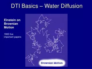
DTI Basics – Water Diffusion. Einstein on Brownian Motion. 1905 five important papers. Why MRI : Detection of Acute Stroke. “Diffusion Weighted Imaging (DWI ) has proven to be the most effective means of detecting early strokes” Lehigh Magnetic Imaging Center. Conventional T 2 WI.

DTI Basics – Water Diffusion
E N D
Presentation Transcript
DTI Basics – Water Diffusion Einstein on Brownian Motion 1905 five important papers
Why MRI : Detection of Acute Stroke “Diffusion Weighted Imaging (DWI) has proven to be the most effective means of detecting early strokes” Lehigh Magnetic Imaging Center Conventional T2 WI DW-EPI Sodium ion pumps fail, water goes in cells and can not diffuse.
Tumor T2 (bright water) DWI (x direction) (T2 (bright water)+(diffusion)) T1 + Gadolinium
1st level of complexity Diffusion Weighted Image X direction • Higher diffusion in X direction lower signal Artifact or Abnormality David Porter - November 2000
Pulse Sequence: Gradient-EchoDiffusion Weighting in X direction Excitation 90o RF G -G Gx EPI (T2) Image Acquisition diffusiongradients Gy Gz
Gradient Coils
y Time RF Gx - Gy Gz x NO DIFFUISION protons (Hydrogen) with DIFFUISION signalloss
Time T2 + diffusion T2 RF Gx - Gy Gz T2 (gradient strength)
DIFFUSION MAPS weak (used to remove spin density, T1, T2, TR, and TE effects) S=S0e(-bDxx) Ln(S) = Ln(S0) – bDxx or Dxx = (Ln(S0) – Ln(S))/b Dxx Most Important image S (T2* EPI + weak Diffusion in X direction) S0(T2* EPI)
2nd Level of complexity DWI : 3 Direction • single-shot EPI diffusion-weighted (DW) images with b = 1000s/mm2 and diffusion gradients applied along three orthogonal directions • Higher diffusion lower signal Dxx Dyy Dzz courtesy of Dr Sorensen, MGH, Boston David Porter - November 2000
PE FE SS A Little More Detail Y Diffusion-Weighting Z Diffusion-Weighting X Diffusion-Weighting y x z GFE GPE GSS RF x y z
X Diffusion-Weighting Z Diffusion-Weighting Y Diffusion Weighting Apparent Diffusion Coefficient ADC (AKA TRACE) used in clinical stroke, tumor, etc ADC = (Dxx + Dyy + Dzz)/3 Orientation independent No directional information (ie direction of greatest diffusion)
3rd level of complexityDiffusion Tensor Imaging Basics • Measures water diffusion in at least 6 directions – we use 12 • Echo-planar imaging (fast acquisition) • Collecting small voxels (1.8x1.8x3mm), scanning takes about 10 minutes
Higher diffusion lower signal • Useful for following white matter tracts
Higher diffusion lower signal Isotropic Anisotropic Adapted from: Beaulieu (2002). NMR in Biomed; 15:435-455
DTI ellipsoidmeasure 6 directions to describe z y no diffusion x
Ellipsoid Image Information available through DTI Tract Pierpaoli and Basser, Toward a Quantitative Assessment of Diffusion Anisotropy, Magn. Reson. Med, 36, 893-906 (1996)
Tractography Superior view color fiber maps Lateral view color fiber maps Zhang & Laidlaw: http://csdl.computer.org/comp/proceedings/vis/2004/8788/00/87880028p.pdf.
MRISC axial cor sag Diffusion Tensor Imaging data for cortical spinal tract on right side blue = superior – inferior fibers green = anterior – posterior fibers red = right – left fibers Note tumor is darker mass on left side of axial slice
DxxDxyDxz DyxDyy Dyz Dzx DzyDzz But what is a diffusion tensor? It is a mathematical description of the ellipsoid. no diffusion xy -xy y-z xz z -xz y-z y z x
Dxx Dxy Dxz Dyx Dyy Dyz Dzx Dzy Dzz Dx’x’ 0 0 0 Dy’y’ 0 0 0 Dz’z’ Whatisdiffusion“Tensor” (D)? difussion gradient direction vector y S=S0exp(-bD) labreferenceframe ellipsoidreferenceframe = (mathematical manipulation) Calculate FA (fractional anisotropy) Fiber track
FA (fractional anisotropy) Information available through DTI Dy’y’ Dx’x’ Dz’z’ FA = ((Dx’x’-Dav)2 + (Dy’y’-Dav)2 + (Dz’z’-Dav)2)0.5 (Dx’x’2+Dy’y’2+Dz’z’2)0.5 s av FA = 0.9 FA = 0
FA + color(largest diffusion direction) red = right – left green = anterior – posterior blue = superior - inferior
Tractography Superior view color fiber maps Lateral view color fiber maps Zhang & Laidlaw: http://csdl.computer.org/comp/proceedings/vis/2004/8788/00/87880028p.pdf.
Signal loss : by intra-voxel phase dispersion At the echo time TE, NMR signal is decayed by, - T2 decay (spin-spin diffusion) - diffusive motion For any set of diff. gradient pulses G G 90 180 echo TE
DTI Scalar Parameters • Trace: Magnitude of diffusion in a voxel. • Increases in damaged white matter • Fractional Anisotropy (FA): Measure of directionally-restricted diffusion. • Decreases in damaged white matter Rosenbloom M, et al. (July 2004). NIAA pubs; http://www.niaaa.nih.gov/publications/arh27-2/146-152.htm
The Diffusion Tensor, D • Diffusion is not equal in all directions (anisotropic). • Use this to probe brain structure! • Represent the diffusion pattern at each point in the brain using an ellipsoid.
Diffusion Vector (Colour) Map • The three magnitudes of the diffusion ellipsoid can be shown using three colours (RGB). • Red = Left – Right • Green = Ant. – Pos. • Blue = Sup. – Inf. • Map of major directions of water movement in the brain.
Fibre Tractography • In principle, the locations of major white matter fibre tracts in the brain can be mapped using the information in the colour map, by “following the arrows.”
Hindered Diffusion (diffusion ellipsoid) without hindrance WILSON with hindrance
Information available through DTI – Orientation of λ1 • Useful for following white matter tracts
Information available through DTI -- Aσ s av • Related to the shape of the ellipsoid • Independent of Dav (normalized) • Zero for a sphere, positive for other shapes • Sensitive to myelination and cortical development
Diffusion Tensor Imaging (As) Normal Adult Brain (A maps)
CELL EXTRA-CELLULAR SPACE FREELY DIFFUSING WATER IN EXTRA-CELLULAR SPACE • Higher diffusion lower signal Tissue Sample A Tissue Sample B Freely Diffusing Water = Dark Larger D Restricted Diffusion = Bright Smaller D
K-space view of the spin echo imaging Ky 1 2 3 . . . . . . . n Kx
PE FE SS Diff. Grad. along different axis Y Diffusion-Weighting Z Diffusion-Weighting X Diffusion-Weighting GFE GPE GSS RF
DTI (Diffusion Tensor Imaging) • single-shot EPI diffusion-weighted (DW) images with b = 1000s/mm2 and diffusion gradients applied along three orthogonal directions • Higher diffusion lower signal courtesy of Dr Sorensen, MGH, Boston David Porter - November 2000
Detection of Acute Stroke “Diffusion Weighted Imaging (DWI) has proven to be the most effective means of detecting early strokes” Lehigh Magnetic Imaging Center Conventional T2 WI DW-EPI Sodium ion pumps fail, water goes in cells and can not diffuse.
Tumor T2 (bright water) DWI (x direction) (T2 (bright water)+diffusion) T1 + Gadolinium
Excitation 90o RF G -G Gx Image Acquisition Gy Gz Do X, Y , and Z at the same time
y z x The Diffusion Tensor, D • Diffusion is not equal in all directions (anisotropic). • Use this to probe brain structure! • Diffusion ellipsoid for each voxel
DTI Scalar Parameters • Trace: The magnitude of diffusion in a voxel. • Fractional Anisotropy (FA): The extent to which diffusion is directionally restricted.