Excitation-Contraction Coupling
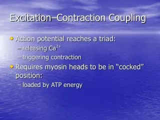
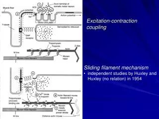
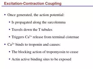
Excitation-Contraction Coupling. Once generated, the action potential: Is propagated along the sarcolemma Travels down the T tubules Triggers Ca 2+ release from terminal cisternae Ca 2+ binds to troponin and causes: The blocking action of tropomyosin to cease

Excitation-Contraction Coupling
E N D
Presentation Transcript
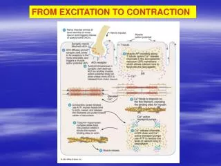
Excitation-Contraction Coupling • Once generated, the action potential: • Is propagated along the sarcolemma • Travels down the T tubules • Triggers Ca2+ release from terminal cisternae • Ca2+ binds to troponin and causes: • The blocking action of tropomyosin to cease • Actin active binding sites to be exposed
Excitation-Contraction Coupling • Myosin cross bridges alternately attach and detach • Thin filaments move toward the center of the sarcomere • Hydrolysis of ATP powers this cycling process • Ca2+ is removed into the SR, tropomyosin blockage is restored, and the muscle fiber relaxes
Excitation-Contraction Coupling Figure 9.9
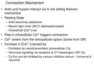
Role of Ionic Calcium (Ca2+) in the Contraction Mechanism • At low intracellular Ca2+ concentration: • Tropomyosin blocks the binding sites on actin • Myosin cross bridges cannot attach to binding sites on actin • The relaxed state of the muscle is enforced Figure 9.10 (a)
Role of Ionic Calcium (Ca2+) in the Contraction Mechanism • At higher intracellular Ca2+ concentrations: • Additional calcium binds to troponin (inactive troponin binds two Ca2+) • Calcium-activated troponin binds an additional two Ca2+ at a separate regulatory site Figure 9.10 (b)
Role of Ionic Calcium (Ca2+) in the Contraction Mechanism • Calcium-activated troponin undergoes a conformational change • This change moves tropomyosin away from actin’s binding sites Figure 9.10 (c)
Role of Ionic Calcium (Ca2+) in the Contraction Mechanism • Myosin head can now bind and cycle • This permits contraction (sliding of the thin filaments by the myosin cross bridges) to begin Figure 9.10 (d)
Sequential Events of Contraction • Cross bridge formation – myosin cross bridge attaches to actin filament • Working (power) stroke – myosin head pivots and pulls actin filament • Cross bridge detachment – ATP attaches to myosin head and the cross bridge detaches • “Cocking” of the myosin head – energy from hydrolysis of ATP cocks the myosin head into the high-energy state
Sequential Events of Contraction Myosin head (high-energy configuration) Myosin cross bridge attaches to the actin myofilament 1 Thin filament ADP and Pi (inorganic phosphate) released Thick filament Working stroke—the myosin head pivots and bends as it pulls on the actin filament, sliding it toward the M line As ATP is split into ADP and Pi, cocking of the myosin head occurs 2 4 Myosin head (low-energy configuration) As new ATP attaches to the myosin head, the cross bridge detaches 3 Figure 9.11
Motor Unit: The Nerve-Muscle Functional Unit Figure 9.12 (a)
Motor Unit: The Nerve-Muscle Functional Unit • A motor unit is a motor neuron and all the muscle fibers it supplies • The number of muscle fibers per motor unit can vary from four to several hundred • Muscles that control fine movements (fingers, eyes) have small motor units
Motor Unit: The Nerve-Muscle Functional Unit • Large weight-bearing muscles (thighs, hips) have large motor units • Muscle fibers from a motor unit are spread throughout the muscle; therefore, contraction of a single motor unit causes weak contraction of the entire muscle
Muscle Twitch • A muscle twitch is the response of a muscle to a single, brief threshold stimulus • The three phases of a muscle twitch are: • Latent period – first few milli-seconds after stimulation when excitation-contraction coupling is taking place
Graded Muscle Responses • Graded muscle responses are: • Variations in the degree of muscle contraction • Required for proper control of skeletal movement • Responses are graded by: • Changing the frequency of stimulation • Changing the strength of the stimulus
Muscle Response to Varying Stimuli • A single stimulus results in a single contractile response – a muscle twitch • Frequently delivered stimuli (muscle does not have time to completely relax) increases contractile force – wave summation Figure 9.14
Muscle Response to Varying Stimuli • More rapidly delivered stimuli result in incomplete tetanus • If stimuli are given quickly enough, complete tetanus results
Treppe: The Staircase Effect • Staircase – increased contraction in response to multiple stimuli of the same strength • Contractions increase because: • There is increasing availability of Ca2+ in the sarcoplasm • Muscle enzyme systems become more efficient because heat is increased as muscle contracts
Muscle Fatigue • Muscle fatigue – the muscle is in a state of physiological inability to contract • Muscle fatigue occurs when: • ATP production fails to keep pace with ATP use • There is a relative deficit of ATP, causing contractures • Lactic acid accumulates in the muscle • Ionic imbalances are present (extracellularpotassium) • Calcium deficiency • Neurotransmitter (ACH) rundown
Muscle Tone • Is the constant, slightly contracted state of all muscles, which does not produce active movements • Keeps the muscles firm, healthy, and ready to respond to stimulus • Essential for maintaining posture (head upright) • Essential for maintaining blood pressure (tone of smooth muscles in walls of blood vessels) • Responding to activation of stretch receptors in muscles and tendons
Muscle Metabolism: Energy for Contraction • ATP is the only source used directly for contractile activity • As soon as available stores of ATP are hydrolyzed (4-6 seconds), they are regenerated by: • The interaction of ADP with creatine phosphate (CP) • Anaerobic glycolysis • Aerobic respiration
Muscle Metabolism: Energy for Contraction Figure 9.18
Muscle Metabolism: Anaerobic Glycolysis • When muscle contractile activity reaches 70% of maximum: • Bulging muscles compress blood vessels • Oxygen delivery is impaired • Pyruvic acid is converted into lactic acid
Muscle Metabolism: Anaerobic Glycolysis • The lactic acid: • Diffuses into the bloodstream • Is picked up and used as fuel by the liver, kidneys, and heart • Is converted back into pyruvic acid by the liver
Oxygen Debt • Vigorous exercise causes dramatic changes in muscle chemistry • For a muscle to return to a resting state: • Oxygen reserves must be replenished • Lactic acid must be converted to pyruvic acid • Glycogen stores must be replaced • ATP and CP reserves must be resynthesized • Oxygen debt – the extra amount of O2 needed for the above restorative processes
Heat Production During Muscle Activity • Only 40% of the energy released in muscle activity is useful as work • The remaining 60% is given off as heat • Dangerous heat levels are prevented by radiation of heat from the skin and sweating
Force of Muscle Contraction • The force of contraction is affected by: • The number of muscle fibers contracting – the more motor fibers in a muscle, the stronger the contraction • The relative size of the muscle – the bulkier the muscle, the greater its strength • Degree of muscle stretch – muscles contract strongest when muscle fibers are 80-120% of their normal resting length
Isometric Contractions Figure 9.17 (b)
Muscle Fiber Type: Functional Characteristics • Speed of contraction – determined by speed in which ATPases split ATP • The two types of fibers are slow and fast • ATP-forming pathways • Oxidative fibers – use aerobic pathways • Glycolytic fibers – use anaerobic glycolysis • These two criteria define three categories – slow oxidative fibers, fast oxidative fibers, and fast glycolytic fibers
Muscle Fiber Type: Speed of Contraction • Slow oxidative fibers contract slowly, have slow acting myosin ATPases, and are fatigue resistant • Fast oxidative fibers contract quickly, have fast myosin ATPases, and have moderate resistance to fatigue • Fast glycolytic fibers contract quickly, have fast myosin ATPases, and are easily fatigued
Smooth Muscle • Composed of spindle-shaped fibers with a diameter of 2-10 m and lengths of several hundred m • Lack the coarse connective tissue sheaths of skeletal muscle, but have fine endomysium • Organized into two layers (longitudinal and circular) of closely apposed fibers • Found in walls of hollow organs (except the heart) • Have essentially the same contractile mechanisms as skeletal muscle
Smooth Muscle Figure 9.24
Peristalsis • When the longitudinal layer contracts, the organ dilates and contracts • When the circular layer contracts, the organ elongates • Peristalsis – alternating contractions and relaxations of smooth muscles that mix and squeeze substances through the lumen of hollow organs
Innervation of Smooth Muscle • Smooth muscle lacks neuromuscular junctions • Innervating nerves have bulbous swellings called varicosities • Varicosities release neurotransmitters into wide synaptic clefts called diffuse junctions
Innervation of Smooth Muscle Figure 9.25
Microscopic Anatomy of Smooth Muscle • SR is less developed than in skeletal muscle and lacks a specific pattern • T tubules are absent • Ca2+ is sequestered in the extracellular space, allowing rapid influx when channels are opened • There are no visible striations and no sarcomeres • Thin and thick filaments are present
Proportion and Organization of Myofilaments in Smooth Muscle • Ratio of thick to thin filaments is much lower than in skeletal muscle • Thick filaments have heads along their entire length • There is no troponin complex • Thick and thin filaments are arranged diagonally, causing smooth muscle to contract in a corkscrew manner
Proportion and Organization of Myofilaments in Smooth Muscle Figure 9.26
Contraction of Smooth Muscle • Whole sheets of smooth muscle exhibit slow, synchronized contraction • They contract in unison, reflecting their electrical coupling with gap junctions • Action potentials are transmitted from cell to cell • Some smooth muscle cells: • Act as pacemakers and set the contractile pace for whole sheets of muscle • Are self-excitatory and depolarize without external stimuli
Contraction Mechanism • Actin and myosin interact according to the sliding filament mechanism • The final trigger for contractions is a rise in intracellular Ca2+ • Ca2+ is released from the SR and from the extracellular space • Ca2+ interacts with calmodulin and myosin light chain kinase to activate myosin
Role of Calcium Ion • Ca2+ binds to calmodulin and activates it • Activated calmodulin activates the kinase enzyme • Activated kinase transfers phosphate from ATP to myosin cross bridges • Phosphorylated cross bridges interact with actin to produce shortening • Smooth muscle relaxes when intracellular Ca2+ levels drop
Special Features of Smooth Muscle Contraction • Unique characteristics of smooth muscle include: • Smooth muscle tone • Slow, prolonged contractile activity • Low energy requirements • Response to stretch
Hyperplasia • Certain smooth muscles can divide and increase their numbers by undergoing hyperplasia • This is shown by estrogen’s effect on the uterus • At puberty, estrogen stimulates the synthesis of more smooth muscle, causing the uterus to grow to adult size • During pregnancy, estrogen stimulates uterine growth to accommodate the increasing size of the growing fetus
Developmental Aspects: Regeneration • Cardiac and skeletal muscle become amitotic, but can lengthen and thicken • Smooth muscle has good regenerative ability
Developmental Aspects: Male and Female • There is a biological basis for greater strength in men than in women • Women’s skeletal muscle makes up 36% of their body mass • Men’s skeletal muscle makes up 42% of their body mass
Developmental Aspects: Age Related • With age, connective tissue increases and muscle fibers decrease • Muscles become stringier and more sinewy • By age 80, 50% of muscle mass is lost (sarcopenia) • Regular exercise can dramatically slow sarcopenia • Aging of the cardiovascular system affects every organ in the body