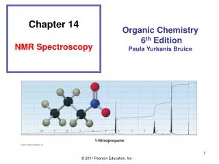
Chapter 14 NMR Spectroscopy
Chapter 14 NMR Spectroscopy. Organic Chemistry 6 th Edition Paula Yurkanis Bruice. Nuclear Magnetic Resonance (NMR) Spectroscopy. Identify the carbon–hydrogen framework of an organic compound. Certain nuclei, such as 1 H, 13 C, 15 N, 19 F, and 31 P, have

Chapter 14 NMR Spectroscopy
E N D
Presentation Transcript
Chapter 14 NMR Spectroscopy Organic Chemistry 6th Edition Paula Yurkanis Bruice
Nuclear Magnetic Resonance (NMR) Spectroscopy Identify the carbon–hydrogen framework of an organic compound Certain nuclei, such as 1H, 13C, 15N, 19F, and 31P, have non-zero value for their spin quantum number; this property allows them to be studied by NMR
The spin state of a nucleus is affected by an applied magnetic field:
The energy difference between the spin states increases with the strength of the applied magnetic field:
absorb DE a-spin states b-spin states release DE Signals detected by NMR
An NMR Spectrometer In pulsed Fourier transform (FT) spectrometers, the magnetic field is held constant, and a radio frequency (rf) pulse of short duration excites all the protons simultaneously
The electrons surrounding a nucleus decrease the effective magnetic field sensed by the nucleus: Beffective = Bo – Blocal
Chemically equivalent protons: protons in the same chemical environment Each set of chemically equivalent protons in a compound gives rise to a signal in an 1H NMR spectrum of that compound:
The Chemical Shift The common scale for chemical shifts = d distance downfield from TMS (Hz) d = operating frequency of the spectrometer (MHz) The reference point of an NMR spectrum is defined by the position of TMS (zero ppm): The chemical shift is a measure of how far the signal is from the reference signal
1H NMR spectrum of 1-bromo-2,2-dimethylpropane The greater the chemical shift, the higher the frequency The chemical shift is independent of the operating frequency of the spectrometer
Protons in electron-poor environments show signals at high frequencies Electron withdrawal causes NMR signals to appear at higher frequency (at larger d values):
Characteristic Values of Chemical Shifts
Diamagnetic Anisotropy The unusual chemical shifts associated with hydrogens bonded to carbons that form p bonds: Thep electrons are freer to move than the s electrons in response to a magnetic field
The protons show signals at higher frequencies because they sense a larger effective magnetic field: benzene
The alkene and aldehyde protons also show signals at higher frequencies: alkene aldehyde
The alkyne proton shows a signal at a lower frequency than it would if the p electrons did not induce a magnetic field: alkyne
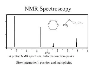
1H NMR spectrum of 1-bromo-2,2-dimethylpropane The area under each signal is proportional to the number of protons giving rise to the signal:
Integration Line The area under each signal is proportional to the number of protons that give rise to that signal The height of each integration step is proportional to the area under a specific signal The integration tells us the relative number of protons that give rise to each signal, not the absolute number
Splitting of the Signals • An 1H NMR signal is split into N + 1 peaks, where N is • the number of equivalent protons bonded to adjacent • carbons • Coupled protons split each other’s signal • The number of peaks in a signal is called the multiplicity • of the signal • The splitting of signals, caused by spin–spin coupling, • occurs when different kinds of protons are close to one • another
It is not the number of protons giving rise to a signal that determines the multiplicity of the signal It is the number of protons bonded to the immediately adjacent carbons that determines the multiplicity a: a triplet b: a quartet c: a singlet
The ways in which the magnetic fields of three protons can be aligned:
Splitting is observed if the protons are separated by no more than three s bonds: Long-range coupling occurs over systems, such as benzene
Triplet: two neighboring protons Quintet: four neighboring protons More Examples of 1H NMR Spectra
Doublet: one neighboring proton Sextet: five neighboring protons Septet: six neighboring protons Triplets: two neighboring protons
The three vinylic protons are at relatively high frequency because of diamagnetic anisotropy
The signals for the Hc, Hd, and He protons overlap because the electronic effect of an ethyl substituent is similar to that of a hydrogen:
The signals for the Ha, Hb, and Hc protons do not overlap because of the strong electron-withdrawing property of the nitro group:
Coupling Constants The coupling constant (J) is the distance between two adjacent peaks of a split NMR signal in hertz: Coupled protons have the same coupling constant
Summary 1. The number of chemical shifts specify the number of proton environments in the compound 2. The chemical shift values specify the nature of the chemical environment: alkyl, alkene, etc. 3. The integration values specify the relative number of protons 4. The splitting specifies the number of neighboring protons 5. The coupling constants specify the orientation of the coupled protons
A Splitting Diagram for a Doublet of Doublets
Complex Splitting JAC = JAB Triplet JAC > JAB Doublet of doublets
The trans coupling constant is greater than the cis coupling constant:
A Splitting Diagram for a Quartet of Triplets
Why is the signal for Ha a quintet rather than a triplet of triplet?
The Difference between a Quartet and a Doublet of Doublets Methylene has three neighbors, appears as a quartet Doublet Doublet
When two different sets of protons split a signal, the multiplicity of the signal is determined by using the N + 1 rule separately for each set of the hydrogens, as long as the coupling constants for the two sets are different When the coupling constants are similar, the multiplicity of a signal can be determined by treating both sets of adjacent hydrogens as though they were equivalent
Replacing one of the enantiotopic hydrogens by a deuterium or any other atom or group other than CH3 or OH forms a chiral molecule: prochiral carbon Ha is the pro-R-hydrogen, whereas Hb is the pro-S-hydrogen; and they are chemically equivalent