Download
1 / 8
80 likes | 117 Vues
Learn about the structure, blood supply, and common issues related to palatine tonsils. Discover how tonsillitis and quinsy are diagnosed and treated effectively. Explore the lymphatic drainage and atrophy process of these lymphoid tissues.

E N D
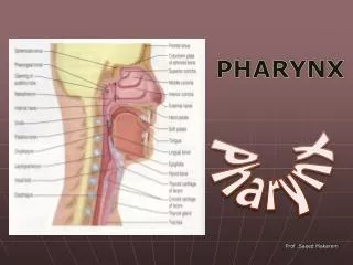
PHARYNX Prepared by/ Ass. Lecturer: Sara Mostafa
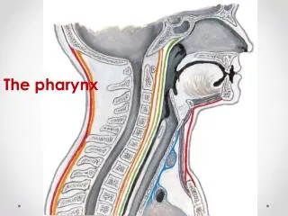
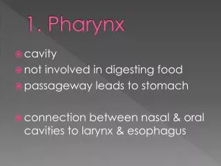
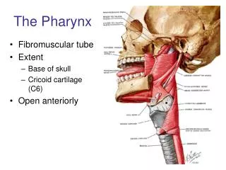
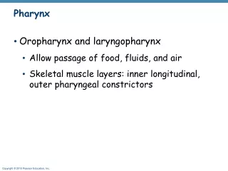
Palatine Tonsils • The palatine tonsils are two masses of lymphoid tissue, each located in the depression on the lateral wall of the oral part of the pharynx between the palatoglossal and palatopharyngeal arches • Each tonsil is covered by mucous membrane, and its free medial surface projects into the pharynx • The surface is pitted by numerous small openings that lead into the tonsillar crypts
Palatine Tonsils • The tonsil is covered on its lateral surface by a fibrous capsule • The capsule is separated from the superior constrictor muscle by loose areolar tissue • The external palatine vein descends from the soft palate in this tissue to join the pharyngeal venous plexus • Lateral to the superior constrictor muscle lie the styloglossus muscle, the loop of the facial artery, and the internal carotid artery
Blood Supply • The tonsillar branch of the facial artery • The veins pierce the superior constrictor muscle and join the external palatine, the pharyngeal, or the facial veins • Lymph drains into the upper deep cervical lymph nodes, just below and behind the angle of the mandible
Tonsils and Tonsilitis • The palatine tonsils reach their maximum normal size in early childhood • After puberty, together with other lymphoid tissues in the body, they gradually atrophy • The palatine tonsils are a common site of infection, producing the characteristic sore throat and pyrexia.
Tonsils and Tonsilitis • The deep cervical lymph node situated below and behind the angle of the mandible, which drains lymph from this organ, is usually enlarged and tender • Recurrent attacks of tonsillitis are best treated by tonsillectomy • After tonsillectomy, the external palatine vein, which lies lateral to the tonsil, may be the source of troublesome postoperative bleeding
Quinsy • A peritonsillar abscess (quinsy) is caused by spread of infection from the palatine tonsil to the loose connective tissue outside the capsule • The nasopharyngeal tonsil or pharyngeal tonsil consists of a collection of lymphoid tissue beneath the epithelium of the roof of the nasal part of the pharynx • Like the palatine tonsil, it is largest in early childhood and starts to atrophy after puberty