Unveiling the Genetic Secrets of DNA: A Molecular Journey
380 likes | 470 Vues
Explore the chemical structure, transformation principles, and genetic significance of DNA, from the pneumococci experiment to Watson and Crick's discovery of the double helix structure. Learn about base pairing, X-ray diffraction, and the tautomeric conversions in this fascinating molecular realm.

Unveiling the Genetic Secrets of DNA: A Molecular Journey
E N D
Presentation Transcript
FCH 532 Lecture 3 Chapter 5: DNA
Figure 1-16 Double-stranded DNA. • Each DNA base is hydrogen bonded to a base on the opposite strand forming a base pair. • A bonds with T and G bonds with C forming complementary strands. Page 18
Figure 5-3 Mechanism of base-catalyzed RNA hydrolysis. Base induced deprotonation of 2’-OH allows nucleophilic attack on the adjacent phosphate group. Results in a cyclic intermediate that can be hydrolyzed into either a 2’ product or 3’ product. Because DNA is resistant to this type of hydrolysis (lack of 2’-OH group) is likely reason to be carrier of genetic info. Page 83
DNA is the transforming principle • Diplococcus pneumoniae is a pneumococcus bacterium that causes pneumonia. • Virulent strains a gelatinous polysaccharide coating that contains binding sites through which it infects cells. • Mutant pneumococci that lack this coating are not virulent (nonpathogenic). • Virulent and nonpathogenic pneumococci are known as S (smooth) and R (rough) respectively. • Experiment: In 1928, Frederick Griffith injected mice with a mixture of live R (nonpathogenic) and heat-killed S (virulent) pneumococci that resulted in the death of most mice. • Dead mice contained live S pneumococci. • The progeny were also S. • Transformation could take place outside the cell by mixing R cells with cell-free extract from R cells. • What is the transforming principle?
Figure 5-4 Pneumococci. Page 83
DNA is the transforming principle • 1944: Avery, MacLeod, and McCarty reported that the transforming principle was DNA. • Results: chemical had all the physical and chemical properties of DNA, no detectable proteins, unaffected by enzymes that hydrolyze proteins and RNA, totally inactivated by enzymes that catalyze the hydrolysis of DNA-therefore, DNA must be the carrier of genetic info. • Eukaryotes can also be transformed by DNA. • Gene for growth hormone (polypeptide) injected into the nuclei of fertilized mouse eggs. • Fertilized mouse eggs implanted into foster mothers resulting in supermice. • Genetically altered known as transgenic.
Figure 5-5 Transgenic mice. Page 84
DNA is the genetic carrier for many bacteriophages • Phage acts like a hypodermic needle full of the transforming principle that is injected into the bacterial host cell. • Tested in 1952 by Hershey and Chase.
Figure 5-6 Bacteriophages attached to the surface of a bacterium. Page 84
Figure 5-7 Diagram of T2 bacteriophage injecting its DNA into an E. coli cell. Page 84
Figure 5-8 The Hershey-Chase experiment. • Used bacteriophage T2 grown on E. coli medium containing 32P and 35S radioisotopes. Radiolabeled the capsid (no P) with 35S and labeled the DNA (no S) with 32P. Page 85
Enol Keto Keto-enol tautomerism Equilibrium between ketone or aldehyde and enol forms. Keto and enol forms are considered tautomers of one another. Carbonyl (C=O) is in rapid equilibrium with enol tautomer that has a double-bonded (C=C) adjacjent to a hydroxyl (OH) group.
Figure 5-9 Some possible tautomeric conversions for bases. Page 87
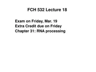
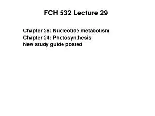
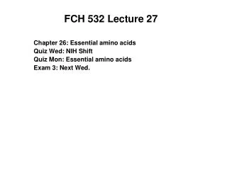
Figure 5-10 X-ray diffraction photograph of a vertically oriented Na+ DNA fiber in the B conformation taken by Rosalind Franklin. • Central X-shaped pattern of spots indicative of helix. • Heavy black arcs on top and bottom correspond to a distance of 3.4 Å • DNA structure repeats every 3.4 Å. Page 87
Double helical DNA • Structure determined by Watson and Crick-suggested the molecular mechanism of heredity. • Tied together several studies: • Chargaff’s rules. A = T and G = C importance was shown. • Correct tautomeric forms of the bases. • Information that DNA is a helical molecule by X-ray diffraction done by Rosalind Franklin. This photograph allowed Watson and Crick to deduce that DNA was helical and the planar aromatic bases form a stack of parallel rings parallel to the fiber axis. • Fibers of DNA assume the B confirmation of DNA (B-DNA). • Based on X-ray diffraction patterns.
Double helical DNA • Consists of 2 polynucleotide strands that wind around a common axis with a right-handed twist • 20 Å diameter double helix. • The strands are antiparallel. • Wrapped around each other and cannot be separated without unwinding the helix. • Bases occupy the core and sugar-phosphate chains are on the outside. • Planes of bases are perpendicular to the helix axis. • Each bases is hydrogen bonded to opposite strand to form a planar base pair (complementary base pair).
Double helical DNA • Ideal B-DNA helix has 10 base pairs (bp) per turn. • Helical twist of 36° per bp. • Aromatic bases have van der Waals thickness of 3.4 Å. • Helix has a pitch (rise per turn) of 34 Å.
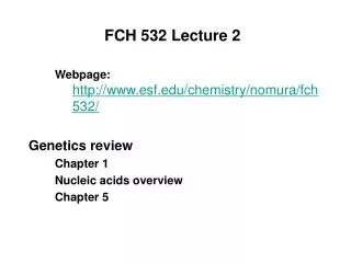
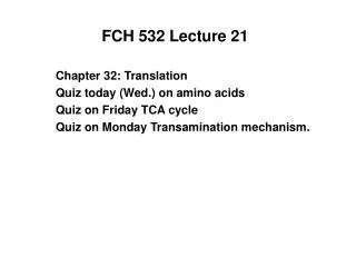
Figure 5-12 Watson-Crick base pairs. Adenine pairs with thymine. Guanine pairs with cytosine. These base pairs are interchangeable in the double helix without altering the positions of the sugar phosphate backbone. The top edge of each base pair is structurally distinct from the bottom edge. The deoxyribose residues are asymmetric. Minor groove exposes the edge from the C1’ atom (open toward bottom) Major groove exposes the opposite edge of each bp. Page 88
Forces in DNA Double Helix • DNA Double helix is stabilized by two types of forces: • H-bonds between complementary bases on opposite strands: • 2 H-bonds in A-T pair • 3 H-bonds in G-C pair • Van der Waals forces and hydrophobic interactions between “stacked bases” • Aromatic bases have p-electrons that interact via attractive Van der Waals forces.
Structure of DNA determines heredity • Watson-Crick bp structure will allow any sequence on one polynucleotide strand as long as the opposite strand has complementary sequence. • Each polynucleotide strand can act as the template for its complementary strand. • In order to replicate, the parental strands must separate so that a complementary daughter strand can be synthesized on each parent strand. • Results in duplex (double-stranded) DNA consisting of one polynucleotide parental strand from the parental molecule and another from the newly synthesized daughter strand. • This is called semi-conservative replication. • Shown by the Meselson-Stahl experiment in 1958. • Increased density of DNA by labeling with 15N and monitored the overall DNA density as a function of growth using equilibrium density gradient centrifugation.
Denaturation and renaturation • Duplex DNA can be heated above a certain temperature to separate the complementary strands into a random coil conformation. • Denaturation is followed by a change in the physical properties of DNA.
Figure 5-14 Schematic representation of the strand separation in duplex DNA resulting from its heat denaturation. Page 90
Denaturation is cooperative • DNA can be monitored by UV absorbance. • When DNA denatures, UV abs is due to aromatic bases and increases compared to the double stranded DNA • Results from disruptions of electronic interactions among nearby bases. • This is called the hyperchromic effect.
Figure 5-15 UV absorbance spectra of native and heat-denatured E. coli DNA. Page 90
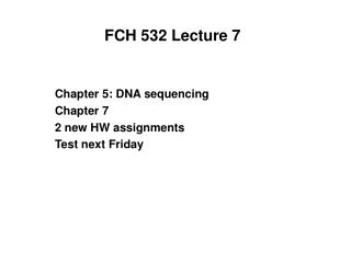
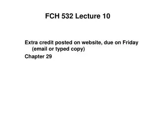
Denaturation is cooperative • The hyperchromic effect takes place over a narrow temperature range. • Indicates that collapse of one part of the DNA duplex will destabilize the rest of the structure (cooperative process). • Melting curves are used to demonstrate the stability of the DNA double helix and determine the melting temperature (Tm) which is the midpoint of a melting curve. • Tm is dependent on the • solvent • concentrations and types of ions • pH • Mole fraction of GC base pairs
Denaturation is cooperative • The hyperchromic effect takes place over a narrow temperature range. • Indicates that collapse of one part of the DNA duplex will destabilize the rest of the structure (cooperative process). • Melting curves are used to demonstrate the stability of the DNA double helix and determine the melting temperature (Tm) which is the midpoint of a melting curve. • Tm is dependent on the • solvent • concentrations and types of ions • pH • Mole fraction of GC base pairs
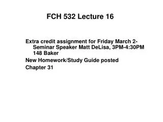
Figure 5-17 Variation of the melting temperatures, Tm, of various DNAs with their G + C content. Page 91
Denatured DNA can be renatured • If a solution of DNA is rapidly cooled below the Tm, the resulting DNA is only partially base paired. • However, if the temperature is maintained at 25 ºC belowe the Tm, the base paired regions will rearrange until DNA completely renatures. • These are called annealing conditions and are important for hybridization of complementary strands of DNA or RNA-DNA hybrid double helices.
Size of DNA molecules • DNA molecules are very large. • Mass can be determined by • ultracentrifugation • length measurements by electron microscopy • Autoradiography-technique in which the position of a radioactive substance in a sample is recorded by exposure to film. • Contour lengths - end to end lengths of stretched out native molecules of DNA. • Genome - complement of genetic information • kb - kilobase pair = 1000 bp
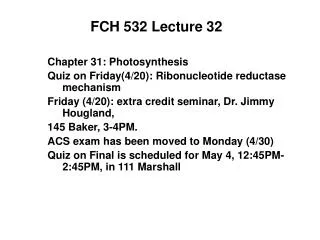
Figure 5-19 Electron micrograph of a T2 bacteriophage and its DNA. Page 91
Figure 5-20 Autoradiograph of Drosophila melanogaster DNA. Page 92
Size of DNA molecules • DNA is highly susceptible to mechanical damage outside of the cell. • Shearing forces generated by ordinary lab techniques can result in shearing of the DNA into small pieces.