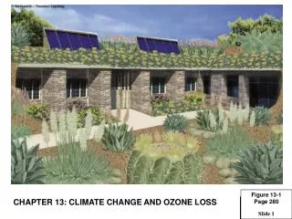
Figure 13-1 Nasal cavities
140 likes | 380 Vues
Figure 13-1 Nasal cavities. Downloaded from: StudentConsult (on 8 August 2011 03:02 PM). © 2005 Elsevier . Figure 13-2 Olfactory mucosa. Downloaded from: StudentConsult (on 8 August 2011 03:02 PM). © 2005 Elsevier . Figure 13-4 Structure of the larynx.

Figure 13-1 Nasal cavities
E N D
Presentation Transcript
Figure 13-1 Nasal cavities Downloaded from: StudentConsult (on 8 August 2011 03:02 PM) © 2005 Elsevier
Figure 13-2 Olfactory mucosa Downloaded from: StudentConsult (on 8 August 2011 03:02 PM) © 2005 Elsevier
Figure 13-4 Structure of the larynx Downloaded from: StudentConsult (on 8 August 2011 03:03 PM) © 2005 Elsevier
Figure 13-5 Structure of the trachea Downloaded from: StudentConsult (on 8 August 2011 03:03 PM) © 2005 Elsevier
Figure 13-6 Segmentation of the intrapulmonary bronchial tree Downloaded from: StudentConsult (on 8 August 2011 03:03 PM) © 2005 Elsevier
Figure 13-7 Histology of the intrapulmonary bronchial tree Downloaded from: StudentConsult (on 8 August 2011 03:03 PM) © 2005 Elsevier
Figure 13-8 Pulmonary acinus Downloaded from: StudentConsult (on 8 August 2011 03:03 PM) © 2005 Elsevier
Figure 13-9 Transition from terminal bronchiole to respiratory bronchiole Downloaded from: StudentConsult (on 8 August 2011 03:03 PM) © 2005 Elsevier
Figure 13-13 Structure and function of Clara cells Downloaded from: StudentConsult (on 8 August 2011 03:03 PM) © 2005 Elsevier
Figure 13-15 Subdivisions of the respiratory bronchiole: Alveolar duct, alveolar sac, and alveoli
Figure 13-19 Type II alveolar cell Downloaded from: StudentConsult (on 8 August 2011 03:03 PM) © 2005 Elsevier