HYPERSENSITIVITY
HYPERSENSITIVITY. Week 15. Hypersensitivity. Definition : Exaggerated or inappropriate response of body's immune system. Causes inflammatory response and tissue damage. Four types. Types I, II, III are antibody mediated. Type IV is T-cell mediated. Type I. Immediate hypersensitivity

HYPERSENSITIVITY
E N D
Presentation Transcript
HYPERSENSITIVITY Week 15
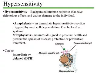
Hypersensitivity • Definition: Exaggerated or inappropriate response of body's immune system. • Causes inflammatory response and tissue damage. • Four types. • Types I, II, III are antibody mediated. • Type IV is T-cell mediated.
Type I • Immediate hypersensitivity • Allergic reaction. Immediately follows antigen contact. • The antigen is classified as an allergen. • “Allergy” is synonymous with Type I. • Family history has a major role. • Atopy: asthma, eczema, hay fever, food allergy, urticaria (hives).
Type I • Levels of circulating IgE to an allergen determine whether an anaphylactic reaction will occur upon re-exposure to the same Ag. • The exact mechanism that leads B cells to produce IgE is not known Mechanisms: • Non-allergic patient • Ag enters body. • IgM produced (primary response). • Second response: IgG produced by plasma cells. Very low levels of IgE produced. • Allergic patient • Very high levels of IgE produced by plasma cells. • IgE associates with two cell types: basophils and mast cells. • These cells have surface receptors for the Fc region of the IgE molecule.
Type I • Non-allergic patient • Many epitopes (antigenic determinants) expressed on cell surfaces in low amounts. • Allergic patient • Also low levels of many different epitopes (idiotypes), but large amount of sites to a particular epitope.
Type I • Note that several IgE's are anchored on surfaces of mast cells and basophils. IgE molecules must be close to one another in order for a response to be generated to antigenic binding. This is the case with allergic patients due to very high levels of IgE. • Mast cells and basophils release histamine and other factors (heparin, chemotactic factors, platelet activating factors). • Histamine causes: smooth muscle contraction, vasodilation, increased vascular permeability. • Normal response is controlled. If out of control anaphylactic response. • If the allergen is injected into circulation (i.e. Not localized) e.g. Insect venom systemic anaphylaxis with dyspnoea, bronchospasm, laryngeal edema and vasodilation sudden drop in BP. • If allergen enters mucous eg pollen, house dust etc. local reaction occurs in respiratory areas. • If allergen in intestinal mucosa eg nuts, strawberries and fish, a mixed reaction occurs including skin rashes and asthma. • The higher individual's IgE level, the greater chances of allergy – strong family association.
Type II • Definition: production of Ab against a self-molecule or against a foreign Ag bound to a cell surface, an infectious agent or inert material damaging reactions (inappropriate host response).
Type II • ADCC: Antibody-dependant cell-mediated cytotoxicity. • In Type II hypersensitivity, antibodies bind to cells or to an antigen adsorbed onto host cells. • Cells involved: neutrophils, eosinophils, monocytes and NK cells. • Examples: incompatible blood transfusion, rhesus-incompatibility, Ab against self-molecules, e.g.: • Thyroid cells (Hashimoto's thyroiditis), kidney cells (Goodpasture's Syndrome), muscle cells (Myesthemia gravis). • Sedormid is a drug that adsorbs on to platelets. Ab directed at drug destroy platelets. • Some infections e.g. salmonella or Mycobacterial infections – endotoxins coat patient's cells, cells destroyed by antibodies.
Type III • Type III (Immune complex) hypersensitivity • Size and form of immune complex depends on how much Ag and Ab are involved. • Large complex formation determined by: • Class of Ab (e.g. IgM much bigger, with multiple binding sites compared to IgG) • Biding strength of antigen. • Sometimes Ag-Ab complex comes out of solution. • Monocytes and macrophages remove large complexes, but don't clear complexes with excess Ag very well. • Neutrophils clear only large complexes.
Type III • If excess Ag present inflammation. This is normal. • If immune complex persists or become trapped in tissues Type III hypersensitivity: • Ischemia develops (capillary networks become blocked) • Arthus' reaction: local reaction. • If an Ag is injected into circulation of a sensitized patient, Ag-Ab complexes deposit in walls of blood vessels redness, swelling, heat, pain (i.e. vasculitis). • Resolves after 24 hrs. (e.g. diabetics reacting to animal-derived insulin).
Type III • Respiratory type: asthma development – approx. 8 hrs later Arthus' reaction in respiratory system. • Usually due to defective working of macrophages, neutrophils or complement; OR system • overloaded with complexes due to continuous large presence of the antigen like blood sepsis. • Triggers mast cells to degranulate. • Neutrophils attracted – release their toxic granules > tissue damage. • Platelet activation factor stimulated microthrombi. • If slight Ag excess – local hypersensitivity in tissues. • If large excess – Ag-Ab complexes spill over into circulation serum sickness • Ag-Ab complexes may also deposit in skin, kidneys and joints (elephantiatis – enormous swellings).
Type IV • Type IV ( Cell mediated or Delayed ) hypersensitivity • Specifically provoked. • Slow to evolve • Involves lymphocytes and macrophages. • T-memory cells of specific antigenic determinant are long lived cells remaining a part of immune system after a primary response. • Circulate through body of sensitized individual. At re-exposure to epitope (presented by APC on MHC molecule). • Proliferation occurs and lymphokines released > attract macrophages; stimulate T-cytotoxic cells (CD8+) > eliminate Ag. • Type IV hypersensitivity occurs when an EXAGGERATED CELL MEDIATED IMMUNE RESPONSE occurs.
Type IV • Examples: • Chronic infectious diseases eg Mycobacteria (TB) and fungi. • Host unable to eliminate antigen continuous release of lymphokines continued accumulation of macrophages cells fuse together – form giant cells. • Macrophages expressing epitope on MHC release more lymphokines tissue damage granuloma • More examples: • Granulomas form against indigestible inorganic materials like silica and talc, • Measles and herpes lesions, • Metals e.g. nickel (in watch straps), • poison ivy, • potassium dichromate in cement, • penicillin. • These substances on their own may not be antigenic; but when combined with protein e.g. in skin: • Langerhans cells take Ag to lymph nodes. T-cells return to entry site to release lymphokines. • Reaction site shows mononuclear infiltrate (lymphocytes and macrophages). • Clinical symptoms : eczema – redness, swelling, vesicles on skin, scaling, exudate.