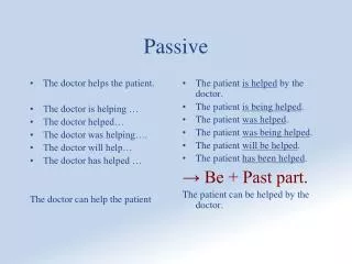

Passive Processes in Membrane Permeation
E N D
Presentation Transcript
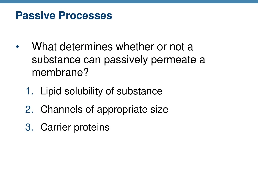
Passive Processes • What determines whether or not a substance can passively permeate a membrane? • Lipid solubility of substance • Channels of appropriate size • Carrier proteins
Passive Processes • Simple diffusion • Carrier-mediated facilitated diffusion • Channel-mediated facilitated diffusion • Osmosis
Passive Processes: Simple Diffusion • Nonpolar lipid-soluble (hydrophobic) substances diffuse directly through the phospholipid bilayer
Extracellular fluid Lipid- soluble solutes Cytoplasm (a) Simple diffusion of fat-soluble molecules directly through the phospholipid bilayer Figure 3.7a
Passive Processes: Facilitated Diffusion • Certain lipophobic molecules (e.g., glucose, amino acids, and ions) use carrier proteins or channel proteins, both of which: • Exhibit specificity (selectivity) • Are saturable; rate is determined by number of carriers or channels • Can be regulated in terms of activity and quantity
Facilitated Diffusion Using Carrier Proteins • Transmembrane integral proteins transport specific polar molecules (e.g., sugars and amino acids) • Binding of substrate causes shape change in carrier
Lipid-insoluble solutes (such as sugars or amino acids) (b) Carrier-mediated facilitated diffusion via a protein carrier specific for one chemical; binding of substrate causes shape change in transport protein Figure 3.7b
Facilitated Diffusion Using Channel Proteins • Aqueous channels formed by transmembrane proteins selectively transport ions or water • Two types: • Leakage channels • Always open • Gated channels • Controlled by chemical or electrical signals
Small lipid- insoluble solutes (c) Channel-mediated facilitated diffusion through a channel protein; mostly ions selected on basis of size and charge Figure 3.7c
Passive Processes: Osmosis • Movement of solvent (water) across a selectively permeable membrane • Water diffuses through plasma membranes: • Through the lipid bilayer • Through water channels called aquaporins (AQPs)
Water molecules Lipid billayer Aquaporin (d) Osmosis, diffusion of a solvent such as water through a specific channel protein (aquaporin) or through the lipid bilayer Figure 3.7d
Passive Processes: Osmosis • Water concentration is determined by solute concentration because solute particles displace water molecules • Osmolarity: The measure of total concentration of solute particles • When solutions of different osmolarity are separated by a membrane, osmosis occurs until equilibrium is reached
(a) Membrane permeable to both solutes and water Solute and water molecules move down their concentration gradients in opposite directions. Fluid volume remains the same in both compartments. Right compartment: Solution with greater osmolarity Both solutions have the same osmolarity: volume unchanged Left compartment: Solution with lower osmolarity H2O Solute Solute molecules (sugar) Membrane Figure 3.8a
(b) Membrane permeable to water, impermeable to solutes Solute molecules are prevented from moving but water moves by osmosis. Volume increases in the compartment with the higher osmolarity. Both solutions have identical osmolarity, but volume of the solution on the right is greater because only water is free to move Left compartment Right compartment H2O Solute molecules (sugar) Membrane Figure 3.8b
Importance of Osmosis • When osmosis occurs, water enters or leaves a cell • Change in cell volume disrupts cell function
Tonicity • Tonicity: The ability of a solution to cause a cell to shrink or swell • Isotonic: A solution with the same solute concentration as that of the cytosol • Hypertonic: A solution having greater solute concentration than that of the cytosol • Hypotonic: A solution having lesser solute concentration than that of the cytosol
(a) Isotonic solutions (b) Hypertonic solutions (c) Hypotonic solutions Cells retain their normal size and shape in isotonic solutions (same solute/water concentration as inside cells; water moves in and out). Cells lose water by osmosis and shrink in a hypertonic solution (contains a higher concentration of solutes than are present inside the cells). Cells take on water by osmosis until they become bloated and burst (lyse) in a hypotonic solution (contains a lower concentration of solutes than are present in cells). Figure 3.9
Summary of Passive Processes • Also see Table 3.1
Cytoplasm • Located between plasma membrane and nucleus • Cytosol • Water with solutes (protein, salts, sugars, etc.) • Cytoplasmic organelles • Metabolic machinery of cell • Inclusions • Granules of glycogen or pigments, lipid droplets, vacuoles, and crystals
Membranous Mitochondria Peroxisomes Lysosomes Endoplasmic reticulum Golgi apparatus Nonmembranous Cytoskeleton Centrioles Ribosomes Cytoplasmic Organelles
Mitochondria • Double-membrane structure with shelflike cristae • Provide most of cell’s ATP via aerobic cellular respiration • Contain their own DNA and RNA
Outer mitochondrial membrane Ribosome Mitochondrial DNA Inner mitochondrial membrane (a) Cristae Matrix (c) Enzymes (b) Figure 3.17
Ribosomes • Granules containing protein and rRNA • Site of protein synthesis • Free ribosomes synthesize soluble proteins • Membrane-bound ribosomes (on rough ER) synthesize proteins to be incorporated into membranes or exported from the cell
Endoplasmic Reticulum (ER) • Interconnected tubes and parallel membranes enclosing cisternae • Continuous with nuclear membrane • Two varieties: • Rough ER • Smooth ER
Smooth ER Nuclear envelope Rough ER Ribosomes (a) Diagrammatic view of smooth and rough ER Figure 3.18a
Rough ER • External surface studded with ribosomes • Manufactures all secreted proteins • Synthesizes membrane integral proteins and phospholipids
Smooth ER • Tubules arranged in a looping network • Enzyme (integral protein) functions: • In the liver—lipid and cholesterol metabolism, breakdown of glycogen, and, along with kidneys, detoxification of drugs, pesticides, and carcinogens • Synthesis of steroid-based hormones • In intestinal cells—absorption, synthesis, and transport of fats • In skeletal and cardiac muscle—storage and release of calcium
Golgi Apparatus • Stacked and flattened membranous sacs • Modifies, concentrates, and packages proteins and lipids • Transport vessels from ER fuse with convex cis face of Golgi apparatus • Proteins then pass through Golgi apparatus to trans face • Secretory vesicles leave trans face of Golgi stack and move to designated parts of cell
1 Protein- containing vesicles pinch off rough ER and migrate to fuse with membranes of Golgi apparatus. Rough ER Phagosome ER membrane Plasma mem- brane Proteins in cisterna Pathway C: Lysosome containing acid hydrolase enzymes 2 Proteins are modified within the Golgi compartments. Vesicle becomes lysosome 3 Proteins are then packaged within different vesicle types, depending on their ultimate destination. Secretory vesicle Pathway B: Vesicle membrane to be incorporated into plasma membrane Golgi apparatus Pathway A: Vesicle contents destined for exocytosis Secretion by exocytosis Extracellular fluid Figure 3.20
Lysosomes • Spherical membranous bags containing digestive enzymes (acid hydrolases) • Digest ingested bacteria, viruses, and toxins • Degrade nonfunctional organelles • Break down and release glycogen • Break down bone to release Ca2+ • Destroy cells in injured or nonuseful tissue (autolysis)