Download
1 / 42
420 likes | 636 Vues
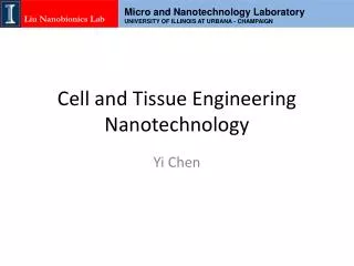
Cell and Tissue Engineering: 3D Effects Lucas Osterbur April 18, 2011 BIOE 506. Overview. Tissue Engineering Background Tissue Engineering in the Third Dimension Motivation Current Fabrication Technologies Effects of 3D – Literature Review Cell-ECM adhesions Cancer Phenotypes

E N D
Cell and Tissue Engineering: 3D Effects Lucas Osterbur April 18, 2011 BIOE 506
Overview • Tissue Engineering Background • Tissue Engineering in the Third Dimension • Motivation • Current Fabrication Technologies • Effects of 3D – Literature Review • Cell-ECM adhesions • Cancer Phenotypes • Stem Cell Differentiation • Personal Work in 3D Tissue Engineering
Tissue Engineering An interdisciplinary field that applies the principles of engineering and life sciences toward the development of biological substitutes that restore, maintain, or improve tissue function • Theoretical solution to wide variety of medical diseases and defects • Organ failure • critical organ shortages • expense • massive immunorepressive responses • Tissue trauma • tissues without regenerative ability • large defect regenerative tissue • Models for human genetic disorders http://biomed.brown.edu/Courses/BI108/BI108_2007_Groups/group12/Homepage.html
Tissue Engineering Demand Surgeries per year related to organ deficiencies Organ Number of US patients Skin >4 million Bone >1 million Heart 750,000 Liver 200,000 Neuromuscular 200,000 Kidney 600,000 http://www.usatoday.com/educate/health/snaps/organ.htm Cheng, J. UIUC, 2010
Tissue Engineering History • 1980 Yannas: Collagen‐GAG scaffold for dermal • regeneration (‘artificial skin’); Integra • • 1985 Wolter/Meyer: 1st use of term, TE; • “Sessile Macrophages Forming Clear Endoethelium-like Membrane on Inside of Successful • • 1993 Langer/Vacanti: Science paper on TE; cells in • matrices for tissue formation in vitro • • 1995 Griffith/Vacanti/Langer: Chondrocytesin • PGA scaffold: the earmouse Zhong et al. Nanomed and Nanobio, 2010, 2, 510-525. Vacanti, C., Langer, R. et al. Plast and Reconstr Surg. 1995, 96, 753.
Tissue Engineering Triad • Scaffold • Porous absorbable material • Regulation of cell functions • Cells • hESCs, MSCs, fibroblasts, chondrocytes, etc. • Autologous, allogenic, xenogenic • Regulators • Chemical – growth factors, small molecules • Genetic – viruses, nucleic acids • Mechanical – mechanical loading, flow conditions Harley, B., UIUC, 2010
Why 3D? • Tissues and organs where cell functions occur are 3D • Our ability to understand formation, function, and pathology often depend on 2D culture or animal models • 2D Cultures • morphology • cell-cell interactions • cell-matrix interactions • differentiation • Animal Models • discrepancies in gene studies • drug therapeutic response • autoimmune disease Yamada et al. Cell, 2007, 130, 601-610
Current 3D Technologies • Multiphoton polymerization Tayalia et al. Adv Mater, 2008. Tayaliaet al. Adv Mater, 2008.
Current 3D Technologies Stachowiak et al. Adv Funct Mat, 2005, 17, 399 – 403.
Current 3D Technologies • Solution Forming Calieri, S. PhD Thesis, Dr. B. Harley
Literature Taking Cell-Matrix Adhesions to the Third Dimension Cukierman, E.; Pankov, R.; Stevens, D.R.; Yamada, K.M. Science, 2001, 294, 1708-1712.
Motivation • Current understanding of cell-matrix adhesions based on in vitro studies of cells and adhesive components • cells display altered morphology and polarity compared to in vivo • Focal Adhesions – integrin based structures • that mediate strong cell-substrate adhesion • integrinαvβ3 j • Paxillin • Vinculin • Focal adhesion kinase • α5and paxillincolocalize in 3D rather than separate to fibrillar and focal sites • “3D Matrix Adhesion” • Fibrillar Adhesions – generate extracellular fibrillars of fibronectin • α5β1 j • tensin
Fibroblast Attachment • 10 minute attachment assay of fibroblasts to various substrates • a.u. = relative number of cells attaching to fibronectin • factor of 6 increase in attachment in control population
Morphology • Threshholded digital images of 4 cells • Only cells distributed on 3D matrix attained elongated in vivo shape within 5 hrs. • Although 2D substrate produced elongated cells after 18 hrs, uncommon branched terminals noted • Only 3D matrix cells significantly altered by treatment with mAb16
Cell Migration and Proliferation • Time lapse video microscopy • 16 paths per treatment method • Migration was promoted by 3D matrix and prohibited by cell treatment with mAb16 • Rates of proliferation more than twice as high in 3D matrix condition
Colocalization in 3D • localization of paxillin, α5integrin, and fibronectin were examined in 5 substrates • cell-derived 3D matrix • tissue-derived 3D matrix • fibronectin • cell-derived 2D matrix • 3D fibronectin Triple colocalization only noted in substrates with a 3D matrix and cell-derived components
Conclusion • Separate localizing of paxillin and integrinswith traditional 2D methods do not adequately describe in vivo fibroblast morphologies • A 3D matrix is necessary for colocalization of adhesion proteins aligned with fibronectin within the matrix • Other components of the cell-derived matrices are also necessary to demonstrate this behavior • Traditional culture methods used to study ECM-Cellular adhesions may not be appropriate for modeling conditions found in vivo
Literature Reversion of Malignant Phenotype of Human Breast Cells in Three-Dimensional Culture and In Vivo by Integrin Blocking Antibodies Weaver, V.M.; Petersen, O.W.; Wang, F.; Larabell, C.A.; Briand, P.; Damsky, C.; Bissel, M.J. The Journal of Cell Biology, 1997, 137, 231-245.
Motivation • About 200,000 new cases of invasive breast cancer will be diagnosed in women in 2011 • In vitro culture may provide a model system to better research the proliferation of cancerous cells and the effects of therapeutic drugs • In breast cancer models, ECM is known to modulate both biochemical and biomechanical signaling events in vivo • ECM signaling pathways may contain suppressor checkpoints the direct and impinge upon cell architecture and tissue • Adherens and other cell-cell junctions are intimately tied into pathways • Final tissue phenotypes may be determined by these pathways and signaling sources • How can 3D culturing methods modify these pathways and the resulting morphogenesis? American Cancer Society
Experimental Design • HMT-3522 breast cancer series used for all experimental cultures • Subline of cells became spontaneously tumorigenic after 238 passages • All non-malignant cells (S-1) derived from passage 50 • Malignant cells (T4-2) derived from passage 238 • Cell line offers unique tool for addressing mechanisms behind malignant conversion in breast cancer cells • Postulate that morphology and behavior of cells can be modified by altering cell-ECM and cell-cell interactions • Commercially available Matrigel used as substrate for 3D matrix • Monolayers grown on thinly coated plastic dishes used for 2D model
Initial Morphology • Only slightly noticeable differences found in growth rate and morphology in 2D • Profound differences evident after just 4 days in 3D system • S-1 formed organized structure similar to those found in benign tumors • T4-2 formed large, loosely organized and invasive colonies • Immunostaining demonstrate basal layer deposition for S-1 cells • Cetenin/Cadherin interaction reduced in T4-2 cells
Integrin Distribution • Both cell types expressed β1, β4, and α6integrins • Distribution patterns radically different • S-1 basally distributed integrins indicative of polarization • T4-2 integrins randomly distributed • Western blot revealed overexpression of β1 and β4 in tumorigenc cells • Malignant behavior a result of integrin changes?
Inhibitory β1 Antibody • S-1 and treated T4-2 exhibit localized nuclei and well organized F-actin • E-cadherins and β-cetenins are colocalized in S-1 and treated T4-2 cells • Control T4-2 cells highly disorganized • Reversible process
Conclusions • The use of related human cell lines, one malignant and one benign allowed for the study of the fundamental role of cell junctions in tissue morphogenesis • Cells produced drastically different results depending on culture in 2D or 3D substrates • T4-2 tumorigenic cells have increased β1integrin expression associated with loss of growth control and pertrubed morphogenesis • A reduction of β1integrin activity is sufficient to revert the tumor phenotype • Malignancy of breast tumor cells could potentially be controlled and restored to normal cell function by experimenting with these interactions
Literature Osteogenic Differentiation of Mouse Embryonic Fibroblast Cells in a Three-Dimensional Self-Assembling Peptide Scaffold Garreta, E.; Genove, E.; Borros, S.; Semino, C.E. Tissue Engineering, 2006, 8, 2215-2227.
Embryonic Stem Cells • Long-Term Self-Renewal: • Can be proliferated for over 100 passages in culture • Keep normal karyotype • Pluripotency: • Human embryonic stem cells have the potential to differentiate into all cell types • How can we best use bioengineering to develop methods for in vitro development of functional tissues from this cell source?
Motivation • Mouse embryonic fibroblast (MEFs) typically employed as a feeder layer for ESCs • Chondrogenic induction of MEFs in high concentration in the presence of bone morphogenic proteins has been found to lead to ossification, providing a model for further bone formation • Cell – matrix adhesions are critical in determining development, differentiation and remodeling of bone • 2D cultures have mostly been used as the substrates for osteoblast-specific differentiation • 3D environments may improve upon the process by attaining the proper niche to simulate the in vivo environment
Loss of Pluripotency • Tagged Oct-4 promoter in mESC cells were monitored throoughout experimental procedure • Loss of fluorescence indicated loss of pluripotency • Confirmation via immunostaining with antibody anti-Oct 4 (red) • Western blot final verification
MEF Osteogenic Differentiation • von Kassa staining (black) confirm mineralization in 3D culture systems • No mineralization found in 2D system • Osteopontin, marker for early stage osteoblast formation, only expressed in 3D immunostaining • 3D culture only system to develop small, elongated phenotype • Phenotype lost when replated in 2D
mESCOsteogenic Differentiation • von Kassa staining confirm mineralization in culture systems • Immunostaining shows presence of ostepontin (red) in cultures • Control staining demonstrates effect of osteogenic pathway • Most notable 2D vs 3D difference in cell proliferation
Conclusion • Experimental procedure let to controlled loss of pluripotency in mESCs • Culture systems were successfully induced into an osteogenic pathway • Dimensionality of culture system is important in determining progress of osteogenic differentiation in mESCs and MEFs • Evidence for MEF cultures are much more supportive of postulation
3D Fabrication • Multiphoton polymerization • Fabrication limitations: • Available materials • Random architecture • Severe processing conditions Tayalia et al. Adv Mater, 2008. Tayaliaet al. Adv Mater, 2008. • Freeze forming • Particle Templating Lee et al. J Mater Chem, 2006. Kim et al. Biomaterials, 2005. Stachowiak et al. Adv Funct Mat, 2005, 17, 399 – 403. Calieri, S. PhD Thesis, Dr. B. Harley
Direct-Write Assembly • Robotic x-y-z control • Layer-by-layer assembly • Pressure driven flow ofcontinuous filament • through deposition nozzle • Materials flexibility (polymers, ceramics, metals…) • Range of architectures and feature sizes from • ~ 1 μm to mm’s 200 mm Lewis and Gratson,,Materials Today (2004).
Direct-Write for Tissue Engineering Polyacrylamide Scaffolds for 3T3 Murine Fibroblasts pHEMA Scaffolds for Rat Hippocampal Culture Barry, R, Shepherd, R. et al. Adv Mat, 2009, 21, 1-4. Hansen Shepherd, J., Parker, S. et al. Adv Funct Mat. 2011, 21, 47-54 HA-Silk Scaffolds for Osteogenic Growth Bio-Inspired Microvascular Networks PhD thesis – S. Parker Wu, W. et al. Adv. Mat. 2011.
Material Requirements • Biocompatible • Biodegradable • Printable Rheology
Material Requirements • Biocompatible • Biodegradable • Printable Rheology • Poly(Hyaluronic Acid) • Natural biopolymer; primary component of ECM • Bioactive material • Proliferation and Survival • Cell Motility • Cell-Cell/Cell-Substrate Adhesion • Long term proliferation in bulk hydrogel Pluripotent hESCs post-20 day culture Gerecht, S. Burdick, J. et al. PNAS, 2005, 104, 11298–11303.
Material Requirements • Biocompatible • Biodegradable • Printable Rheology • Hyaluronidase • Natural enzyme to degrade pHA • Well defined kinetics • Degradation of pHAin vivo K.P. Vercruysse et al. J Chromatogr B, 1994, 6, 179-190
Material System • Biocompatible • Biodegradable • Printable Rheology • Ink Composition • high and low MW methacrylated HA • PEGDA • Irgacure • Water/glycerol solvent • Shear thinning gel • allows for nozzle deposition • without clogging • G’ > G” for spanning features
Hyaluronic Scaffolds 100 µm 200 µm • Scaffolds successfully printed with hyaluronic acid ink via direct-write assembly • Printing and UV curing carried out simultaneously • Feature sizes can be varied systematically
Cell Growth Day 1 Day 3 Day 5 Day 7 Day 9 Day 11 with YijieGeng, Dr. Fei Wang
Conclusion Cell and tissue engineering are moving into the third dimension as researchers seek to investigate cell behavior in the most biomimetic environment possible. As new technologies and methods make this a reality, new cell activities related to differentiation, morphogenesis, and cell adhesions are being discovered that continue to widen the gap between 2D and 3D milieus. The field has an exciting and practically limitless future, and the push from in vitro to in vivo on the lab bench will be an integral part how the progression unfolds.