Download
1 / 72
720 likes | 1.1k Vues
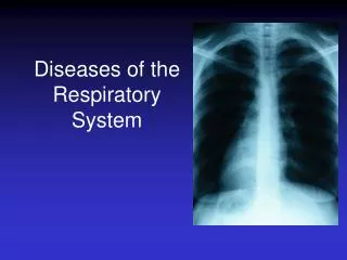
Infection Diseases of Respiratory System in Children. 上海交通大学医学院附属新华医院儿科 鲍一笑. Introduction. High Morbidity Rate High Mortality Rate. Acute and Chronic Infection Rheumatic Disease Pleural Disease Foreign Body of Airway Neoplasm Congenital Anomalies.

E N D
Infection Diseases of Respiratory System in Children 上海交通大学医学院附属新华医院儿科 鲍一笑
Introduction • High Morbidity Rate • High Mortality Rate Acute and Chronic Infection Rheumatic Disease Pleural Disease Foreign Body of Airway Neoplasm Congenital Anomalies Each year, respiratory infection diseases cause about 15 million deaths among children younger than age 5 year through the world. This is a significant cause of mortality in childhood. Pediatric pulmonary infection accounts for about 63.89% of all hospitalizations of children, in which 44.6 percent are pneumonia.
Anatomy and Physiology Venting, Warming, Humidification and conditioning Upper respiratory tract: nose, paranasal sinuses pharynx, eustachian tube, epiglottis, larynx Cricoid cartilage Lower respiratory tract:trachea, bronchi, bronchioles, alveolus ventilation
Nasal cavity is short and narrow More vascular Nasal mucosa Is soft Short Nasal passages, nasolacrimal duct and eustachian tube Significance :These characters make nasal cavity easy to become hyperemia, edema, and congestion which will induce infection. Local infection can spread to nearby organs and tissues easily and cause dyspnea, hoarseness and apnea. Anatomy and Physiology Upper respiratory tract
Anatomy and Physiology Narrowed airway Soft mucous menbrane More vascular Softer and more compliant pulmonary alveoli sIgA on Respiratory Mucosa alveolar surfactant Lower respiratory tract Small amounts Clinical significance: Easy to become hyperemia, edema, and congestion which will induce infection Complication: Pulmonary emphysema and atelectasis
Anatomy and Physiology The younger the child The quicker the frequency The less regular the rhythm Vital capacity (VC) Tidal volume Total lung capacity (TLC) Small Respiratory frequency and rhythm : The respiratory frequency is inversely related to age. ⑴ neonate : 40~50 bpm;6~12mo: 30-35 bpm; 1-3yr : 25~30 bpm;4~9 yr : 20-25bpm; 8-14yr :18~20 bpm。 (2) Some young infants present with irregular rhythm or apnea due to immature respiratory center.
Anatomy and Physiology • Thoracic cage The thorax is barrel shaped. The ribs are in horrizontal position which are almost perpendicular to the spinal column. The location of diaphragm is oppositely superior, which make the size of thoracic cavity decrease, and the size of lung increase. • Respiratory immune function The specific and nonspecific immune function are poor.
Acute Upper Respiratory Infection Acute Upper Respiratory Tract Infection AURI commonly called “common cold”
Introduction • The common cold is the most common pediatric disease and accounts for 80-90% proportion of visit to clinic. • Local infection may spread to nearby organs and tissues which will likely to cause otitis media, conjunctivitis, lymphadenitis, lymphadenitis and pneumonia. • Bronchial asthma, nephritis, myocarditis, measles and pertussis may also follow AURI
Etiology 90% of AURI are caused by viral infection Rhinovirus Echo virus Coxsackievirus Parainfluenza Influenza Adenovirus RSV(Respiratory Syncytial Virus)
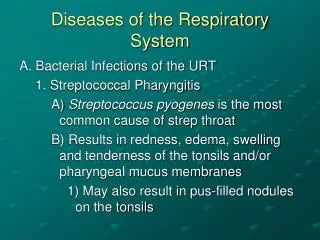
Bacteria Pneumococcus Moraxelle catarrhalis Haemophilus influenzae Staphylococcus aureus
Others Mycoplasma Chlamydia Other Microorganisms
Clinical Manifestation • Mild symptom • Nasal congestion, rhinorrhea, • sneezing, sore throat • Severe symptom • High fever, convulsion, • anorexia,frequency cough
Physical Sign • The pharynx is red • Retropharyngeal folliculosis • Erythematous enlarged tonsils • Enlarged lymph nodes • Enterovirus illnesses may be associated with a wide variety of skin rashes
Two Special Type Herpangina • Coxsackievirus A • Most often occurs in summer and autumn • More often in infants(0-3 yr of age) • Characterized by sudden onset of fever, sore throat and dysphagia • Characteristic lesions, present on the posterior pharynx, are discrete vesicles and ulcers • Duration of illness is usually 7 days
Pharyngoconjunctival Fever • Occurs typically with type 3,7 adenovirus • Most often occurs in spring and summer • Children (>3 yr ) more often affected • Features include: A high temperature that lasts 4–5 days, pharyngitis, conjunctivitis, cervical lymphadenopathy, and rhinitis. • Duration of illness is usually 1-2 weeks
Complication Otitis media Cervical lymphadenitis Bronchitis Pneumonia Septicemia Viral Infection → Viral Myocarditis Viral Encephalitis Bacterial Infections(streptococcus)) → Acute NephritisRheumatic Fever
Diagnosis Clinical manifestations Symptoms and sighs
Differential diagnosis • The differential diagnosis of the URl includes other acute infectious disease. • In patient with febrile convulsion, central nervous system Infections should also considered. • Patients with abdominal pain may have acute abdomen.
Difference Between Mesenteric Lymphadenitis and Acute appendicitis
Prophylaxis • Increase outdoor activities. • Improve physical fitness. • Enhance immunity function. • Patients in collective institutions should be isolated.
Treatment • General treatment Etiological treatment Anti-virus:Ribavirin Avoid the abuse of antibiotics • Symptomatic treatment Severe nasal obstruction Irritability-restlessness High fever Pharyngeal portion ulcer Conjunctivitis
Summary • Upper respiratory infection is the most common disease in childhood • most of which are caused by viral infections. • The severity of clinical manifestations is related to age of the patients. • Infants present mild local symptoms and severe systemic symptoms, while older children present on the contrary. • A stuffy, congested nose may exist in infants younger than 3 months of age. • Treatment for the common cold should be mainly symptomatic. Antibiotics should not be used unless in those young, infant patients which are suspected to complicate bacterial infections.
Acute Bronchitis • Acute bronchitis is inflammation of the tracheobronchial epithelium . • Trachea is usually involved,so acute bronchitis is also called ‘acute tracheobronchitis’. • Acute bronchitis is commonly secondary to an acute viral infection, or just one manifestation of acute infectious disease.
Etiology • Infectious factors:viral, bacterial or other pathogen infections • Characters of respiratory tract of infants: The mucous become edema and hyperemia which make the bronchus narrower when inflammation. • Other factors:immunodeficiency, nutritional diseases, specific body constitution.
Clinical Manifestation • Begins as an URI • Cough is a significant signsnonproductive cough→ productive • The systemic symptoms is usually serve in infants including fever, vomiting and diarrhea • Medical examination: Respiratory rudeness Diffuse or scattered rales No dyspnea • CXR : may be normal or thickening lung markings
Summary Acute bronchitis is an inflammation of the major conducting airways within the lung which caused by viral or bacteria, and is most often in infants. Cough is the most significant clinical manifestation. Fever, vomiting and diarrhea are frequent in infants. Respiratory sounds are rough and scattered rales are heard on auscultation.Radiographic examination of the chest may show a mild increase in bronchovascular markings.Antibiotics are indicated if a bacterial infection of the airway is suspected or proven. Corticosteroids are recommended in severe cases.
Acute Pneumonia • Pneumonia is an inflammation of the parenchyma of the lungs. • Most cases of pneumonia are caused by microorgnanisms, but there are several noninfectious causes, which include aspiration of food or gastric acid, foreign bodies and so on.
Epidemiology • Season of onset • Age of onset • Morbidity rate • Mortality rate
Category Classified according to the infecting organism: Viral pneumonia, bacterial Pneumonia, mycoplasma Pneumonia. Classified according to Pathology: Bronchopneumonia, lobar pneumonia,interstitial pneumonia. Classified according to duration of disease: Acute pneumonia(<1 mo), persistent pneumonia(1-3 mo) and chronic pneumonia(> 3mo). Classified according to severity of disease: Mild pneumonia and severe pneumonia.
Etiology Streptococcus pneumoniae, Haemophilus influenzae, Staphylococcus aureus,Escherichia coli, Pseudomonas pyocyanea Bacteria Viruses Respiratory Syncytial Viruses, adenovirus, influenza, parainfluenza Incidence rate of Chlamydia pneumoniae and Mycoplasma pneumoniae are increasing recent years. others
Inducement Patients with the following problems are particularly predisposed to this disease: Age More often in infants Disease Malnutrition, Congenital heart disease, Immunodeficiency disease Environment The recidence is wetness, stuffiness and crowding.
Pathology • Hyperemia, edema and inflammatory infiltration of lung tissues • Alveolar exudate • Patchy Inflammation focus, and consolidation • Atelectasis and emphysema of lung
ClinicalManifestion cough Fever four pneumonia symptoms Rales tachypnea
Severe Pneumonia Apart from the general features of bronchopneumonia, severe pneumonia also present with systemic toxic symptoms in respiratory system, circulatory system, nervous system and digestive system.
Extrapulmoanry presentations Intracranial hypertension Encephaledema Nervous system Myocarditis, heart failure Microcirculation disturbance Circulatory system Gastrointestinal dysfunction, enteroplegia Alimentary tract hemorrhage Digestive system Mixed acidosis, dehydration Hyponatremia Water-Electrolyte Balance
Myocardial failure • Suddenly onset of tachypnea, R>60 bpm, increasedpulmonary rales. • Tachycardia that can not be explained by high fever or tachypnea, HR>180 bpm • Irritability and cyanosis • Gallop rhythm or dull heart sound , distension of jugular vein and enlarged cardiac • Increased liver with tenderness, > 1.5cm. • Oliguria or anuria that present with edema of eyelid or lower extremities.
Complication • Empyema of pleura Purulent pneumothorax Bullae of lung • Others:Septicemia Purulent pericarditis
Laboratory Examination • Peripheral blood examination White cell count CRP (C-reactive protein) Nitroblue tetrazolium test • Etiological examination Bacteriological examination : Bacterial culture Virological examination: Viral isolation Examinationof mycoplasma: Specific immunity examination
Lobular pneumonia (Bronchopneumonia) • Pathogen Streptococcus pneumoniae Haemophilus influenzae • Pathology Pathological changes such as hyperemia and edemaof bronchiolar wall, exudation of pulmonary lobule, and bronchiolar obstruction are scatteredsurround bronchus. • Clinical manifestation Hyperpyrexia, cough, tachypnea and dyspnea More common in infants, aged people and weak people
Chest radiographic findings in bronchopneumonia • Increase lung markings • Diffuse bilateral Patchy infiltrates and consolidation scattered throughout both lungs • Atelectasis, hyperinflation, • bullae of lungand pyothorax
Chest radiographic findings in bronchopneumonia Frontal views: Patchy infiltrates and consolidation at the inner zone and middle zone of bilateral lower lobes, with or without hyperinflation
Segmental atelectasis Frontal views: It is a segmental atelectasis at the right superior lobe. The transversa fissure is displaced toward the airless lobe. There is a sector high density shadow with the apex toward the hilum of lung. The diaphragm is elevated and the mediastinum is shifted to the side of involvement.
Lobar pneumonia • Pathogen: maily streptococcus pneumoniae • Pathology : inflammtion infiltrates throughout a whole lobe or segment of the lung. • Main clinical manifestation: • More common in adolescence, rare in young children. • Hyperpyrexia, cough, and rusty sputum • X-ray findings Change after changes of clinical symptoms.
Lobar pneumonia at middle lobe of right lung Frontal views: A consolidation within the transverse fissure and oblique fissure can be seen at the middle lobe of right lung,
Bronchiolitis • viral disease, RSV (85%). • aged 2-6 months. • airway obstruction is due to pathological changes include swelling and distension of bronchioles, secretions blockage.
Clinical Manifestation • expiratory wheezing • tachypnea, nasal flaring • Cyanosis • fine rales • emphysema • The duration of illness is 4 ~ 7 days
Chest radiographic findings • Hyperexpansion is commonly present • Peribronchial cuffing • Increased interstitial markings • Patchy infiltrates