Download
1 / 18
590 likes | 2.35k Vues
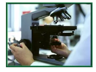
USE AND CARE OF THE MICROSCOPE. LECTURE 1. MICROSCOPY. Light Microscopy: any microscope that uses visible light to observe specimens Compound Light Microscopy: utilizes a series of lenses uses visible light as the source of illumination allows you to observe a specimen as a magnified image

E N D
USE AND CARE OF THE MICROSCOPE LECTURE 1
MICROSCOPY • Light Microscopy: any microscope that uses visible light to observe specimens • Compound Light Microscopy: • utilizes a series of lenses • uses visible light as the source of illumination • allows you to observe a specimen as a magnified image • Resolution=0.2 um • Brightfield Illumination: visualization of dark objects in a bright field
Fine adjustment Coarse adjustment
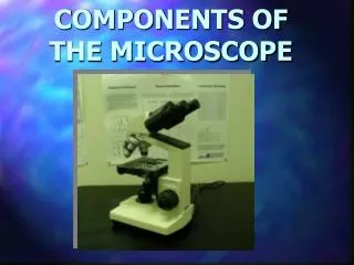
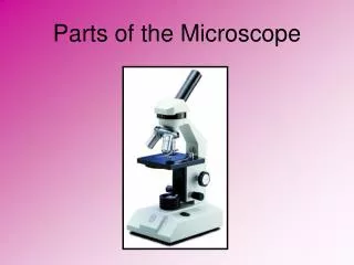
Key Terms • Illuminator: light source • Condenser: series of lenses that direct light through the specimen of interest • Objective lens: first magnification, lens located closest to the specimen, usually 4 different objective lenses (scanning, 10X, 40X, 100X) • Ocular lens: located in the eyepiece, last magnification lens • Coarse adjustment: used with low-power objectives (scanning and 10X) • Fine adjustment: used with high-power objectives (40X and 100X)
Key Concepts • Total Magnification: Multiply the objective lens magnification by the ocular lens (usually 10X)magnification • Resolution/Resolving Power: Ability of the lens to distinguish fine detail and structure or the ability to distinguish two points a specified distance apart. Compound Microscope = 0.2 um. Shorter the wavelength, the greater the resolution. • Oil Immersion: Same effect of increasing the objective lens, therefore it improves the resolving power of the lens • Refractive Index: The measure of the light bending ability of a medium (Ex:Water). Therefore, specimens must be made to contrast their medium. • Use of Stains, Dyes
Other Types of Microscopy • Darkfield:Specimen is light against a dark background, used to examine live organisms • Phase-Contrast: uses special condenser to enhance intracellular details of specimen can be visualized in living organisms/ takes advantage of differences in refractive indexes of cell structures • Differential Interference Contrast (DIC):Similar to Phase-Contrast, higher resolution, image appears 3-D • Fluorescence: Ability of specimens to absorb short wavelengths of light (ultraviolet) and give off long wavelengths of light (visible). Used in diagnostic procedures called fluorescent antibody technique • Confocal: specimen is stained w/ fluorescent dye, computer process the image-uses laser light to illuminate one plane of a specimen at a time. Higher resolution, clear images
Electron Microscopy • Electron Microscopy: • Used for specimens smaller than 0.2 um • Electrons used instead of a beam of light • Higher Resolution due to shorter wavelengths of electrons (~100,000X shorter than visible light) • Types of Electron Microscopes: • Transmission • Scanning
TEM • Transmission Electron Microscopy (TEM): • A beam of electrons is passed through an ultra-thin section of specimen • electromagnetic lenses are used to direct the beam • a copper mesh grid is used to view the sample • a projector lens is used to focus the image on a fluorescent screen • Resolving Power =2.5 nm • Total Magnification =10,000-100,000X • best for virus particles, flagella and proteins • resulting photo is black and white, no 3-D
SEM • Scanning Electron Microscopy (SEM): • Uses an electromagnetic lens to direct electrons over the surface of a specimen • Results in a 3-D image • Resolving Power= 20 nm • Total Magnification = 1,000-10,000X • Best for observing the surfaces of microorganisms
IMPORTANT TIPS TO REMEMBER: • 1-OCULAR AND OBJECTIVE LENSES SHOULD ONLY BE CLEANED WITH LENS PAPER (NOT BIBULOUS PAPER) ALWAYS CLEAN THEM BEFORE AND AFTER USE • 2-OIL SHOULD ONLY BE USED WITH 100X OBJECTIVE • 3-RETURN MICROSCOPES ON SCANNING OBJECTIVE WITH STAGE UP AND SLIDE CENTERED • 4-ALWAYS HANDLE AND CARRY YOUR MICROSCOPE USING THE BASE AND ARM