Quantification of Body Skin Aging with Multiple Parameters
Explore the nature of skin changes with age, their impact on perception, and quantification methods. Factors such as elasticity, hydration, and chromophores play key roles in skin aging. Discover how measuring multiple parameters at various body sites enhances understanding. References to studies on skin aging mechanisms and effects of stressors like UV exposure are provided.

Quantification of Body Skin Aging with Multiple Parameters
E N D
Presentation Transcript
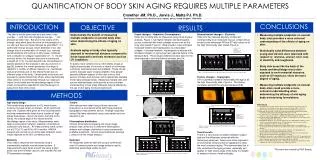
QUANTIFICATION OF BODY SKIN AGING REQUIRES MULTIPLE PARAMETERS Crowther JM. Ph.D., Jarvis J., Matts PJ. Ph.D. P&G Beauty, Rusham Park, Whitehall Lane, Egham, Surrey, United Kingdom, TW20 9NW CONCLUSIONS INTRODUCTION OBJECTIVE RESULTS “ My skin is not the same now as it was when I was younger…”, and “Our skin changes as we age….”, are phrases we often hear. But what is the nature of this change, what effect does this have on the perception of our skin and how can these changes be quantified? It is well known that as we age certain elements of our skin change due to a combination of intrinsic (e.g. reduced dermal elasticity and build up of fatty tissue, combined with a reduction in collagen regeneration) [1, 11, 10] and extrinsic factors (for example exposure to smoke and sunlight) [2, 4, 5]. Current research has concentrated on specific aspects of the changes in skin as a function of age [3], however the overall perception of change is related to a number of different aspects (i.e. mechanical, textural and tonal) and on the effects these have on different areas of the body. Certain parts of the body are exposed to greater stress than others, either mechanically (near joints) or environmentally through sun exposure (backs of the hands and the décolletage region of the chest) and would be expected to show these signs of aging more than other areas. Visual changes – Hydration, Chromophores Older skin is visibly drier as measured using visual dryness analysis, Figure 1, has higher melanin and haemoglobin scores, combined with lower collagen scores over all the body sites tested, Figure 2. Note a lower L value indicates increased melanin and haemoglobin, but decreased collagen. Increased melanin, haemoglobin combined with decrease collagen on aging has been reported before in the literature [6,7]. The strongest differences were seen for the sites exposed to both mechanical stresses and UV. Biomechanical changes – Elasticity Older skin has reduced elasticity component corresponding to an increased viscous component as shown by the decreased R5 and R7 ratio values at all the high stress body sites tested, Figure 3. Demonstrate the benefit of measuring multiple endpoints on several body sites when determining the effects of aging on skin. Evaluate aging on body sites typically exposed to mechanical stresses compared to those exposed to extrinsic stressors such as UV irradiation. • Measuring multiple endpoints on several body sites provides a more coherent understanding of the effects of aging on skin. • Statistically valid differences between young and old skin were observed with respect to: dryness, uneven color, loss of elasticity and roughness. • Body skin areas like the back of the hands and décolletage most often exposed to environmental stressors, such as UV exposure, show the most degree of aging. • Measuring multiple endpoints on several body sites could provide a more coherent understanding when determining the efficacy of anti-aging body moisturizing formulations. • References • 1. Sorg O, Kuenzli S, Kaya G, Saurat J-H. Proposed mechanisms of action for retinoid derivatives in the treatment of skin aging, J Cosmet Dermatol 2005; 4: 237-244. • 2. Leyden JJ. Clinical features of aging skin, Br J Dermatol 1990; 122 (suppl 33): 1-3. • 3. Zimbler MS, Kokoska MS, Thomas JR. Anatomy and pathophysiology of facial aging, Facial Rejuvenation Nonsurgical Modalities 2001; 9(2): 179-187. • 4. Yin L, Moria A, Tsuji T. Skin premature aging induced by tobacco smoking: the objective evidence of skin replica analysis, J Dermatol Sci 2001; 27 (suppl 1): S26-S31. • 5. Kennedy C, Bastiaens MT, Bajdik CD, et al. Effect of smoking and sun on the aging of skin, J Invest Dermatol 2003; 120: 548-554. • 6. Hillebrand GG, Miyamoto K, Schnell B, Ichihashi M, et al. Quantitative evaluation of skin condition in an epidemological survey of females living in northern versus southern Japan, J Dermatol Sci 2001; 27 (suppl 1): S42-S52. • 7. Akazaki S, Nakagawa H, Kazama H, et al. Age-related changes in skin wrinkles as assessed by a novel three-dimensional morphometric analysis, Br J Dematol 2002; 147: 689-695. • 8. Li L, Mac-Mary S, Marsaut D, et al. Age-related changes in skin topography and microcirculation, Arch Dermatol Res 2006; 297: 412-416. • 9. Lagarde JM, Rouvrais C, Black D. Topography and anisotropy of the skin surface with aging, Skin Research and Technology 2005; 11: 110-119. • 10. Ghadially R, Brown BE, Sequeira-Martin SM, Feingold KR, Elias PM. The aged epidermal permeability barrier. Structural, functional, and lipid biochemical abnormalities in humans and a senescent murine model, J Clin Invest 1995; 95(5): 2281-90. • 11. Rogers J, Harding CR, Mayo A, Banks J, Rawlings AV. Stratum corneum lipids: the effect of aging & the seasons, Arch Dermatol Res 1996; 288: 765-770. * demonstrates a significant difference from the 20-30 year age group with p<0.05 * * * * * * * * * * To build a more coherent story of the effects of age on highly stressed parts of the body, a variety of techniques commonly used techniques are available. These can be used to evaluate signs of aging related to how people perceive different aspects of their skin (colour, tone, texture, firmness and dryness) and to specifically evaluate more highly stressed areas. Measuring multiple sites with various methodologies could provide a more coherent understanding of how these attributes may be affected by the use of anti-aging moisturizing products. Figure 3. Skin elasticity values for the different body sites as a function of age and sample images. Visual dryness images from young and old skin 20-30 years old 50-60 years old Texture changes – Topography Skin roughness increases measurably with age at all the high stress body sites, Figure 4. This finding correlates well with the reported literature [8,9]. * * * * * demonstrates a significant difference from the 20-30 year age group with p<0.05 * METHODS Figure 1. Visual dry skin values for the different body sites as a function of age and sample images. Age study design Two female study populations (n=12) were chosen covering the age ranges of interest - 20-30 and 50-60 years old. Subjects were given 20 mins acclimatization and had measurements taken from a number of ‘high stress’ body areas – above the knees, the back of the hands, the outside edge of the elbows and the décolletage region of the chest. Measurements were taken covering skin hydration, elasticity, chromophore distribution and texture. All measurements were carried out at 21ºC±1ºC and 50%±10% humidity. ANOVA analysis was carried out on all the data collected, and p values reported at the 95% confidence level. Elasticity Visco-elastic response was measured using a commercially available vacuum based system. 3 measurements were taken at each site using a 2mm probe size and 100mbar vacuum, and resultant R5 and R7 parameters averaged. Texture Skin replicas were taken using silicone resin and analysed on a commercial white light fringe projection topographic measurements device. Average roughness values (Ra) were calculated using a star pattern and are reported in µm. Chromophore distribution Contact chromophore mapping and visual image analysis was carried out to determine haemoglobin, melanin and collagen distributions using commercially available equipment. Scores are presented as average L values from the image analysis. Hydration An image was taken at each site using a commercial near UV camera system and image analysis used to determine percentage visible dry skin. Typical topographic skin images from young and old skin 20-30 years old 50-60 years old * * * * * demonstrates a significant difference from the 20-30 year age group with p<0.05 * Melanin maps from young and old skin 20-30 years old 50-60 years old * * Figure 4. Average roughness values for the different body sites as a function of age and sample roughness images. * demonstrates a significant difference from the 20-30 year age group with p<0.05 * * * Overall results There is a very strong correlation between subject age and the differences observed with the techniques employed. Areas more often exposed to environmental stressors like UV appeared to show the most consistent aging. This demonstrates that to fully understand the aging process, especially as it applies to ‘high stress’ areas of the body, it is helpful to employ a variety of methodologies. * * * * * * * This work was funded by P&G Beauty Figure 2. Melanin, Haemoglobin and Collagen values for the different body sites as a function of age and sample melanin maps.