EYE
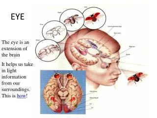
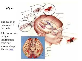
EYE. CONJUNCTIVA. Transparent mucous membrane lining the eyelids and covering anterior surface of eyeball except cornea Richly innervated Vascular. Lacrimal Apparatus. Tears with bactericidal enzyme flow across the eyeball, wash away foreign particles, and help with diffusion of O2 & CO2.

EYE
E N D
Presentation Transcript
CONJUNCTIVA • Transparent mucous membrane lining the eyelids and covering anterior surface of eyeball except cornea • Richly innervated • Vascular
Lacrimal Apparatus • Tears with bactericidal enzyme flow across the eyeball, wash away foreign particles, and help with diffusion of O2 & CO2.
Extrinsic Eyes Muscles trochlea • Innervated by cranial nerves III, IV and VI • 4 rectus muscles move eye up, down, left & right • superior & inferior oblique are complicated
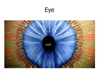
The Tunics of the Eyeball • Fibrous layer (tunica fibrosa) = sclera and cornea • Vascular layer (tunica vasculosa) = choroid, ciliary body & iris • Internal layer (tunica interna) = retina and optic nerve
OPTICAL COMPONENTS • Transparent structures that refract (bend) light rays to focus them on the retina 1. Cornea - covers anterior surface of eyeball 2. Aqueous humor - clear serous fluid located between lens and cornea 3. Lens - suspended by ring of suspensory ligaments 4. Vitreous humor - jelly-like located between the lens and retina
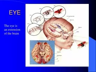
NEURAL COMPONENTS 1. Retina - out growth of diencephalon - pressed against rear of eyeball by vitreous body 2. Optic nerve - attached to retina at the optic disc
IMAGE FORMATION 1.Light passes from an object through the lens 2. A tiny inverted image forms on the retina • Pupillary constrictor (smooth muscle) in iris narrows the pupil in bright light • Pupillary dilator (radial & myoepithelial) widens the pupil in dim light
Refraction • Bending of light rays occurs when light passes through substance with different refractive index at any angle other than 90 degrees • refractive index of air is arbitrarily set to n = 1 • refractive index of cornea is n = 1.38 • refractive index of lens is n = 1.40 • Cornea refracts light more than lens does • lens fine-tunes the image as shift focus between near and distant objects
ACCOMMODATION • Allows the lens to focus on close objects 1. Contraction of ciliary muscle relaxes suspensory ligaments 2. Lens becomes more convex in shape 3. Light is refracted more strongly & focused onto retina
EYE DEFECTS • Hyperopia - farsightedness - due to short eyeball - correct with convex lenses • Myopia - nearsightedness due to long eyeball - correct with concave lenses
Retinal Cells • Posterior layer of retina is pigmented epithelium - absorbs excess light & prevents reflections • Photoreceptors are anterior to epithelium 1. Rod cells - allow night vision - outer segment has a stack of membranous discs containing rhodopsin (pigment) 2.Cone cells - allow color vision in bright light - outer segment tapers to a point
Non-receptor Retinal Cells • Bipolar cells (1st order neurons) • synapse on ganglion cells • moderate convergence occurs • Ganglion cells (2nd order neurons) • axons form optic nerve • great convergence occurs • Horizontal & amacrine cells form connections between other cells
VISUAL PIGMENTS 1. Rhodopsin (visual purple) - in rod cells - consists of opsin (protein) & retinal (vitamin A derivative) 2. Photopsin (iodopsin) - in cones - 3 kinds of cones absorb different wavelengths of light to produce color vision
PHOTOCHEMICAL REACTION IN RODS • Rhodopsin absorbs light & converts from a bent shape (cis-retinal) to a straight (trans-retinal) form that dissociates from opsin (bleaching) • 50% of rhodopsin is regenerated 5 minutes after bleaching occurs
Color Vision • Cones permit color vision • Cones are named for absorption peaks of photopsins • blue cones peak sensitivity at 420 nm • green cones peak at 531 nm • red cones peak at 558 nm (orange-yellow) • Perception of color is based on mixture of nerve signals • Color blindness is hereditary lack of one photopsin • Red-green color blindness (sex-linked recessive trait found in 8% of males) occurs if an individual lacks either red or green cones
Rods vs. Cones RODS CONES