EYE
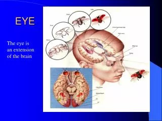
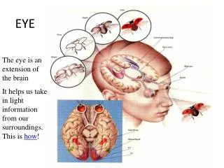
EYE. The eye is an extension of the brain It helps us take in light information from our surroundings. This is how !. Eye brain proximity. Can you see : the optic nerve bundle? Spinal cord?. What are the parts of the eye?.

EYE
E N D
Presentation Transcript
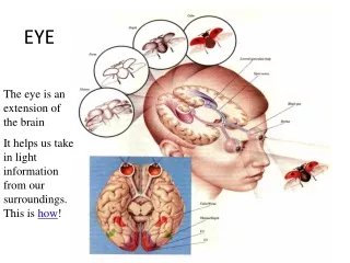
EYE The eye is an extension of the brain It helps us take in light information from our surroundings. This is how!
Eye brain proximity • Can you see : • the optic nerve bundle? • Spinal cord?
What are the parts of the eye? • Let’s use a diagram to help us get familiar with the parts and pronounce them correctly!
Cornea Cornea- transparent membrane that covers iris and pupil. Focuses light on the retina.
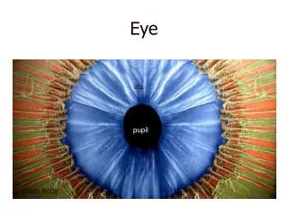
Pupil, Iris, Sclera Pupil- hole passes different amounts of light Iris- colored part of the eye that controls the size of the pupil Sclera- “whites” of the eye that forms outer covering
Aqueous Humor, Ciliary Muscle, Lens Aqueous Humor- nutritious fluid between the iris and the cornea. As we age, it breaks down and we begin to see “floaters” Ciliary Muscles- produces aqueous humor and controls lens shape Lens- Bends light rays to the back of the eye –retina. Elastic so change in shape allows for focus on objects close up or far away
Retina • Full of light receptors which are sensitive to: • Cones- Colour • Rods- Light levels • Massive blood supply is also needed Blind Spot- site of optic nerve connection This is Ms. Vikingson’s retina!!
Vitreous Humor, Blind Spot, Optic Nerve, Vitreous Humor- transparent jelly-like fluid that fills the eye and refracts light Optic Nerve- sends messages picked up by retina to the brain Blind Spot- area where optic nerve attaches. No retina is located there so information cannot be picked up; “Blind”
Choroid Layer, Tapetum lucidum Choroid Layer- lies between the sclera and the retina it provides the blood supply to the eye. Tapetum lucidum- iridescent film under the retina that provides animals with “night vision”
Cross section Sclera Choroid Layer Taptem lucidum You must know: • Lens • Cornea • Aqueous humor • Pupil • Iris • Ciliary Muscle • Sclera • Vitreous Humor • Retina • Blind Spot • Choroid Layer • Optic Nerve • Tapetum Lucidum Blind Spot Ciliary
Eye Dissection • Before we go over the dissection, let’s review the parts of the eye and their function
Select a place to make an incision of the sclera midway between the cornea and optic nerve. Use the point of a surgical scissors to make a small cut through the sclera. Fluid should ooze out of the eyeball when you have cut deeply enough.
Arrange the two hemispheres of the eye as you see in the photograph. • Observe the semi-fluid vitreous humor that fills the central cavity of the eye. It is transparent in the living eye but might be cloudy in the preserved specimen
The retina lines the the posterior cavity of the eye and extends forward to the ciliary body. Use your probe to lift and pull the retina back from the underlying choroid layer. • Notice that the retina is only firmly attached to the choroid at one place. This region is the optic disc or blind spot.
Remove the lens and place against newspaper to see that it is a magnifier!
When the lens is removed, an opening, allowing light to enter the eye is seen. This opening, the pupil is located in the center of the iris. Note the oblong shape of the sheep pupil, in humans the pupil is circular. • The back side of the iris can be seen just above the pointer in the photograph.
Can you identify the parts? You will need to to get credit during the lab
1. Cornea 2. Sclera 3. Optic Nerve 4. Iris 5. Pupil 6. Ora Serrata (you do not have to know this structure!) 7. Ciliary Body 8. Choroid 9. Tapetum Lucidum 10. Retina 11. Lens 12. Vitreous Humor
Clean up! • Once all eye parts have been located and signed off by your teacher, it is time to clean up! • Clean off all instruments on your paper towel and put them away • Wrap up eye and all eye parts inside your paper towel • Remove gloves around paper towel for easy disposal • Place items into garbage can