Download
1 / 33
360 likes | 930 Vues
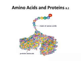
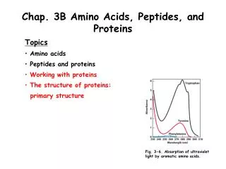
Chapter 25: Amino Acids, Peptides, and Proteins. monomer unit: -amino acids Biopolymer: the monomeric amino acids are linked through an amide bond (the carboxylic acids of one AA with the -amino group of a second) peptide (< 50 amino acids) protein (> 50 amino acids).

E N D
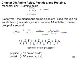
Chapter 25: Amino Acids, Peptides, and Proteins. monomer unit: -amino acids Biopolymer: the monomeric amino acids are linked through an amide bond (the carboxylic acids of one AA with the -amino group of a second) peptide (< 50 amino acids) protein (> 50 amino acids) Peptide or protein (polypeptide)
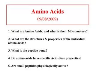
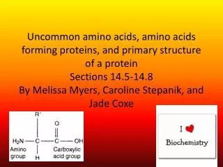
25.1: Classification of Amino Acids. AA’s are classified according to the location of the amino group. There are 20 genetically encoded-amino acids found in peptides and proteins 19 are primary amines, 1 (proline) is a secondary amine 19 are “chiral”, 1 (glycine) is achiral; the natural configuration of the -carbon is L.
-Amino acids are classified by the properties of their sidechains. Nonpolar: Polar but non-ionizable:
Acidic: Basic: 25.2: Stereochemistry of Amino Acids: The natural configuration of the -carbon is L. D-Amino acids are found in the cell walls of bacteria. The D-amino acids are not genetically encoded, but derived from the epimerization of L-isomers (Ch. 25.6).
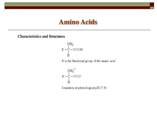
25.3: Acid-Base Behavior of Amino Acids. Amino acids exist as a zwitterion: a dipolar ion having both a formal positive and formal negative charge (overall charge neutral). pKa ~ 5 pKa ~ 9 Amino acids are amphoteric: they can react as either an acid or a base. Ammonium ion acts as an acid, the carboxylate as a base. Isoelectric point (pI): The pH at which the amino acid exists largely in a neutral, zwitterionic form (influenced by the nature of the sidechain) Table 25.2 & 25.3 (p. 1126)
pKa1 + pKa2 2 pI = pI = 6.0 pKa1 + pKa3 2 pI = pI = 2.7 pKa2 + pKa3 2 pI = pI = 9.7 pKax + pKay 2 pI =
Electrophoresis: separation of polar compounds based on their mobility through a solid support. The separation is based on charge (pI) or molecular mass.
25.4: Synthesis of Amino Acids: Ch. 20.15 Strecker Synthesis: recall reductive amination (Ch. 20.10) and Cyanohydrin formation (Ch. 17.7) Amidomalonate Synthesis: recall the malonic acid synthesis (Ch. 20.11)
25.5: Reactions of Amino Acids. Amino acids will undergo reactions characteristic of the amino (amide formation) and carboxylic acid (ester formation) groups. 25.6: Some Biochemical Reactions of Amino Acids. Many enzymes involved in amino acid biosynthesis, metabolism and catabolism are pyridoxal phosphate (vitamin B6) dependent. (please read)
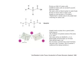
25.7: Peptides. Proteins and peptides are polymers made up of amino acid units (residues) that are linked together through the formation of amide bonds (peptide bonds) from the amino group of one residue and the carboxylate of a second residue By convention, peptide sequences are written left to right from the N-terminus to the C-terminus backbone
The amide (peptide) bond has C=N double bond character due to resonance resulting in a planar geometry restricts rotations resistant to hydrolysis The N-H bond of one amide linkage can form a hydrogen bond with the C=O of another. N-O distance 2.85 - 3.20 Å optimal N-H-O angle is 180 ° Disulfide bonds: the thiol groups of cysteine can be oxidizedto form disulfides (Cys-S-S-Cys)
Epidermal Growth Factor (EGF): the miracle of mother’s spit 53 amino acid, 3 disulfide linkages 1986 Nobel Prize in Medicine or Physiology : Stanley Cohen Rita Levi-Montalcini
25.8: Introduction to Peptide Structure Determination. Protein Structure: primary (1°) structure: the amino acid sequence secondary (2°): frequently occurring substructures or folds tertiary (3°): three-dimensional arrangement of all atoms in a single polypeptide chain quaternary (4°): overall organization of non-covalently linked subunits of a functional protein. Determine the amino acids present and their relative ratios Cleave the peptide or protein into smaller peptide fragments and determine their sequences Cleave the peptide or protein by another method and determine their sequences. Align the sequences of the peptide fragments from the two methods
E-A-Y-L-V-C-G-E-R F-V-N-Q-H-L-F-S-H-L-K G-C-F-L-P-K L-G-A F-V-N-Q-H-L-F S-H-L-K-E-A-Y L-V-C-G-E-R-G-C-F L-P-K-L-G-A F-V-N-Q-H-L-F F-V-N-Q-H-L-F-S-H-L-K S-H-L-K-E-A-Y E-A-Y-L-V-C-G-E-R L-V-C-G-E-R-G-C-F G-C-F-L-P-K L-P-K-L-G-A L-G-A F-V-N-Q-H-L-F-S-H-L-K-E-A-Y-L-V-C-G-E-R-G-C-F-L-P-K-L-G-A
25.9: Amino Acid Analysis. automated method to determine the amino acid content of a peptide or protein Reaction of primary amines with ninhydrin Enzymatic digestion peptide -or- protein [H] reduce any disulfide bonds individual amino acids -or- H3O+, liquid chromatography derivatize w/ ninhydrin Detected w/ UV-vis Different amino acids have different chromatographic mobilities (retention times) 1972 Nobel Prize in Chemistry William Stein Stanford Moore
25.10: Partial Hydrolysis of Peptides. Acidic hydrolysis of peptides cleave the amide bonds indiscriminately. Proteases (peptidases): Enzymes that catalyzed the hydrolysis of the amide bonds of peptides and proteins. Enzymatic cleavage of peptides and proteins at defined sites: • trypsin: cleaves at the C-terminal side of basic residues, Arg, Lys but not His • chymotrypsin: cleaves at the C-terminal side of aromatic residues Phe, Tyr, Trp
Trypsin and chymotrypsin are endopeptidases Carboxypeptidase: Cleaves the amide bond of the C-terminal amino acid (exopeptidase) 25.11: End Group Analysis. The C-terminal AA is identified by treating with peptide with carboxypeptidase, then analyzing by liquid chormatography (AA Analysis). N-labeling: The peptide is first treated with 1-fluoro-2,4-dinitro benzene (Sanger’s reagent), which selectively reacts with the N-terminal amino group. The peptide is then hydrolyzed to their amino acids and the N-terminal amino acid identified as its N-(2,4-dinitrophenyl) derivative (DNP).
25.12: Insulin. Insulin has two peptide chains (the A chain has 21 amino acids and the B chain has 30 amino acids) held together by two disulfide linkages. (please read) Pepsin: cleaves at the C-terminal side of Phe, Tyr, Leu; but not at Val or Ala. Pepsin cleavage Trypsin cleavage H3O+ cleavage
25.13: The Edman Degradation and Automated Peptide Sequencing. Chemical method for the sequential cleavage and identification of the amino acids of a peptide, one at a time starting from the N-terminus. Reagent: Ph-N=C=S, phenylisothiocyanate (PITC) -1 peptide with a new N-terminal amino acid (repeat degradation cycle) N-phenylthiohydantoin: separated by liquid chromatography (based of the R group) and detected by UV-vis
Peptide sequencing by Edman degradation: • Cycle the pH to control the cleavage of the N-terminal amino acid by PITC. • Monitor the appearance of the new N-phenylthiohydantoin for each cycle. • Good for peptides up to ~ 25 amino acids long. • Longer peptides and proteins must be cut into smaller fragments before Edman sequencing. Tandem mass spectrometry has largely replaced Edman degradation for peptide sequencing (Fig. 25.10, p. 1146) 25.14: The Strategy for Peptide Synthesis: Chemical synthesis of peptide: 1. Solution phase synthesis 2. Solid-phase synthesis
The need for protecting groups Orthogonal protecting group strategy: the carboxylate protecting group must be stable to the reaction conditions for the removal of the -amino protecting group and ( vice versa)
25.15: Amino Group Protection. The -amino group is protectedas a carbamate. 25.16: Carboxyl Group Protection. Protected as a benzyl ester; removed by hydrogenolysis
25.17: Peptide Bond Formation. Amide formation from the reaction of an amine with a carboxylic acid is slow. Amide bond formation (peptide coupling) can be accelerated if the carboxylic acid is activated. Reagent: dicyclohexylcarbodiimide (DCC)
• In order to practically synthesize peptides and proteins, time consuming purifications steps must be avoided until the very end of the synthesis. • Large excesses of reagents are used to drive reactions forward and accelerate the rate of reactions. • How are the excess reagents and by-products from the reaction, which will interfere with subsequent coupling steps, removed without a purification step? 25.18: Solid-Phase Peptide Synthesis: The Merrifield Method. Peptides and proteins up to ~ 100 residues long are synthesized on a solid, insoluble, polymer support. Purification is conveniently accomplished after each step by a simple wash and filtration.
The solid support (Merrifield resin): polystyrene polymer Solid-phase peptide synthesis
Ribonuclease A- 124 amino acids, catalyzes the hydrolysis of RNA Solid-phase synthesis of RNase A: Synthetic RNase A: 78 % activity 0.4 mg was synthesized 2.9 % overall yield average yield ~ 97% per coupling step His-119 A His-12 A LYS GLU THR ALA ALA ALA LYS PHE GLU ARG GLN HIS MET ASP SER SER THR SER ALA ALA SER SER SER ASN TYR CYS ASN GLN MET MET LYS SER ARG ASN LEU THR LYS ASP ARG CYS LYS PRO VAL ASN THR PHE VAL HIS GLU SER LEU ALA ASP VAL GLN ALA VAL CYS SER GLN LYS ASN VAL ALA CYS LYS ASN GLY GLN THR ASN CYS TYR GLN SER TYR SER THR MET SER ILE THR ASP CYS ARG GLU THR GLY SER SER LYS TYR PRO ASN CYS ALA TYR LYS THR THR GLN ALA ASN LYS HIS ILE ILE VAL ALA CYS GLU GLY ASN PRO TYR VAL PRO VAL HIS PHE ASP ALA SER VAL His-12 B His-119 B pdb code: 1AFL R. Bruce Merrifield, Rockefeller University, 1984 Nobel Prize in Chemistry: “for his development of methodology for chemical synthesis on a solid matrix.”
25.19: Secondary Structures of Peptides and Proteins. -sheet: Two or more extended peptide chain, in which the amide backbones are associated by hydrogen bonded N C anti-parallel loop or turn N C C N C N parallel N C N C crossover N C N C
-helix: 3.6 amino acids per coil, 5.4 Å C N 3.6 AA 5.4 Å
myoglobin pdb code: 1WLA Bacteriorhodopsin pdb code: 1AP9 Anti-parallel -sheets of lectin pdb code: 2LAL Parallel -sheets carbonic anhydrase pdb code: 1QRM
25.20: Tertiary Structure of polypeptides and Proteins. Fibrous. Polypeptides strands that “bundle” to form elongated fibrous assemblies; insoluble. Globular. Proteins that fold into a “spherical” conformation. Hydrophobic effect. Proteins will fold so that hydrophobic amino acids are on the inside (shielded from water) and hydrophilic amino acids are on the outside (exposed to water) Pro • Ile • Lys • Tyr • Leu • Glu • Phe • Ile • Ser • Asp • Ala • Ile • Ile • His •Val • His • Ser • Lys
Enzymes: proteins that catalyze biochemical reactions. • by bringing the reactive atoms together in the optimal geometry for the reaction. • lowering the activation energy (G‡) by stabilizing the transition state and/or high energy intermediate. • many enzymes use the functional groups of the amino acid sidechain to carry out the reactions Proteases (peptidases):catalyzes the hydrolysis of peptide bonds Four classes of proteases: Serine (trypsin): aspartate-histidine-serine Aspartyl (HIV protease, renin): two aspartates Cysteine (papain, caspase): histidine-cysteine Metallo (Zn2+) (carboxypeptidase, ACE): glutamate
Mechanism of carboxylpeptidase, metalloprotease (p. 1162) Mechanism of a serine protease (trypsin, chymotrypsin):
25.21: Coenzymes. Some reactions require additional organic molecules or metal ions. These are referred to as cofactors or coenzymes. 25.22: Protein Quaternary Structure: Hemoglobin. (please read) 25.23: G-Coupled Protein Receptors. (please read)