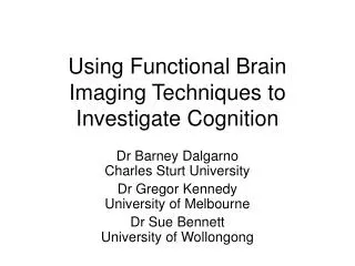
Using Functional Brain Imaging Techniques to Investigate Cognition
240 likes | 432 Vues
Using Functional Brain Imaging Techniques to Investigate Cognition. Dr Barney Dalgarno Charles Sturt University Dr Gregor Kennedy University of Melbourne Dr Sue Bennett University of Wollongong. Our Overall Research Question.

Using Functional Brain Imaging Techniques to Investigate Cognition
E N D
Presentation Transcript
Using Functional Brain Imaging Techniques to Investigate Cognition Dr Barney DalgarnoCharles Sturt University Dr Gregor KennedyUniversity of Melbourne Dr Sue BennettUniversity of Wollongong
Our Overall Research Question How does interactivity in multimedia environments impact on learners' cognitive processes and subsequent learning outcomes?
Brain Imaging Methods • Functional Magnetic Resonance Imaging (fMRI) • Positron Emission Tomography (PET) • Single Photon Emission Computed Tomography (SPECT) • Electro-Encephalography (EEG)
PET, SPECT • The participant is first injected with a tracer radionuclide. • Tracer leaves a temporary signature in the active regions of the brain during a subsequent task. • Tracer detected by PET or SPECT scanning. • Short half-life water-based radionuclides: • Scanning at regular intervals during task (eg. every couple of minutes) • Long half-life glucose based radionuclides: • Scanning after completion of task (eg. half an hour) • Participant can carry out tasks on a computer outside of the scanner and then undergo scanning to measure overall activation patterns during the session.
EEG • Detection of signals in cortical areas of the brain via sensors placed on surface of the scalp • Very high temporal fidelity (eg. many readings per second) • Low spatial resolution (eg. 20 sensors)
fMRI • The participant lies with their head and upper body inside a scanner with their head still. • A projected computer image is viewed via a mirror above the participant’s head. • Interaction occurs using simple hand-held buttons or a special purpose mouse. • Headphones and a microphone can be used. • Regional (3D) fluctuations in magnetic fields are measured, which correlate with blood flow and in turn brain activation. • The cost is about $1000 per participant.
Our Study: Questions • Is there a detectable difference in the overall brain activation between users of a simulation-based and a tutorial-based multimedia learning resource? • If so, does this difference explain predicted differences in learning outcomes? • Is brain activation during identified interactive episodes consistent with accepted theory?
Hypotheses (Cognition) • Simulation learners will experience: • Deeper elaborative processing and cognitive organisation, as they regularly draw on current understanding in decision making towards a task goal. • Greater degrees of knowledge construction, reflection and refinement through regular feedback following actions within the environment.
Hypotheses (Brain Activation) Elaborative processing and cognitive organisation: • The hippocampus, the dorsolateral prefrontal cortex(DLPFC) and the ventrolateral prefrontal cortex(VLPFC) have been associated with elaborative processing and cognitive organisation. Feedback-based learning, performance monitoring and control: • Feedback based learning has been found to result in activation of the basal ganglia, including the striatum and the caudate nucleus along with areas of the prefrontal cortex (PFC) and the posterior frontomedian cortex (pFMC). • Tasks requiring choices to be made have been found to activate the anterior cingulate cortex (ACC) and where conflicting options are available, pFMC activation has been found. • Tasks requiring error detection and monitoring of activity have been found to also activate the ACC. • Cognitive control mechanisms have been associated with the mid DLPFC.
Methods • Eight participants, four conditions • 2 resource types (tutorial, simulation) X 2 content areas (climate change, blood alcohol). • Traditional measures: pencil and paper questionnaires, observation, video stimulated recall and audit trails) • fMRI: activation scan every 3 seconds • Analysis: • Overall activation (beyond ‘baseline’) in the two resource types • Activation during interactive tasks (event-related methods)
Problems Encountered • Limitations on interactivity due to availability of MRI compatible input devices • Activation from motor tasks in simulation could confound the results. • Visual differences between conditions could confound the results. • Regular baseline conditions problematic during holistic task.
Addressing these Problems • Compromises in design of multimedia resources. • Identical screen layouts in tutorial and simulation. • Simulation structure: i. plan manipulations and predict outcomesii. carry out manipulations and simulate; iii. review feedback; iv. rest screen (baseline). • 3-button interface device.
Analysis and Interpretation • Examples from the final pilot participant
Will our results be conclusive? • Definitive findings may require a degree of certainty from the literature, in identifying the functional characteristics of each brain area. • It is not yet possible to associate activation in a particular brain area with a single cognitive function. • However, we believe that it is possible to make conclusions about the degree to which activation patterns are consistent or inconsistent with certain theories.
Where to Next • fMRI analysis and triangulation with test, questionnaire and interview data • Larger non-imaging study
Acknowledgements • fMRI advice: • Dr David Abbott and other staff from the Brain Research Institute • PET advice: • Dr Graeme O’Keefe, Austin Department of PET and Geoff Currie, CSU Nuclear Medicine • Theoretical advice: • Professor Terry Bossemaier, CSU • Programming advice and audit trail coding: • Dr Terry Judd, University of Melbourne • Global warming model assistance: • Professor David Battisti, University of Washington and Dr Andrew Hall, Charles Sturt University • Blood alcohol concentration model development: • Dr Michael Lew, University of Melbourne • Funding: • CSU Small Grant • University of Wollongong Research Centre for Interactive Learning Environments (RILE) Seed Grant