Download
1 / 1
10 likes | 190 Vues
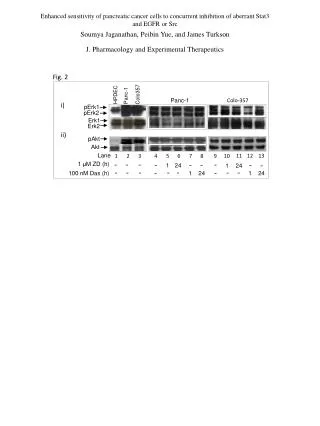
Progesterone receptor membrane component 1 (PGRMC1) and its role in the establishment and progression of female cancers Melissa L. McCallum 1 , Cindy A. Pru 1 , Hannah Balash, Bo R. Rueda 2 , John J. Peluso 3 , James K. Pru 1

E N D
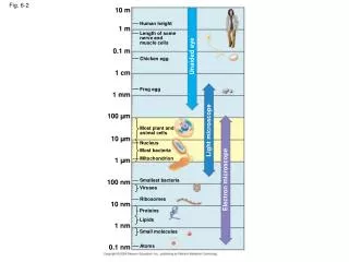
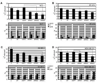
Progesterone receptor membrane component 1 (PGRMC1) and its role in the establishment and progression of female cancers Melissa L. McCallum1, Cindy A. Pru1, Hannah Balash, Bo R. Rueda2, John J. Peluso3, James K. Pru1 Department of Animal Sciences1, Washington State University, Pullman, WA 99164; Vincent Center for Reproductive Biology2, Harvard Medical School, Boston, MA 02114; Department of Cell Biology3; University of Connecticut Health Center, Farmington, CT 06030 INTRODUCTION The second leading cause of death among women in the United States is cancer. Approximately 40,000 women die of breast cancer annually, and another 40,000 are diagnosed with endometrial cancer. Often, the way endometrial cancers are treated is by completely removing the female reproductive tract, leading to infertility. Many cancers involving the female reproductive system depend on hormone regulation. In order to investigate the role of progesterone in cancer, studies in our lab are focused on a novel membrane progestin receptor called progesterone receptor membrane component 1, or PGRMC1. We are working to determine if PGRMC1 is important in the growth and chemoresistance of tumors. Here, a simple subtraction approach was taken in which PGRMC1 either remained intact in a cell population or was depleted. The PGRMC1 knockdown cells were produced by injecting a cell colony with a Lentivirus that eliminated the PGRMC1 gene. Two cell lines, the MDA-MB-231 breast cancer cells and the Ishikawa (IKLV) endometrial cancer cells were used for the experiments. Microarray analysis was performed and this resulted in differential regulation of many genes that was verified primarily by reverse transcriptase polymerase chain reaction (RT-PCR). Western blot analysis was also used to validate microarray data for some genes. Then human xenograft tumors were developed in immunocompromised mouse models. Six mice were injected intraperitoneally (into the body cavity) with five million GFP-labeled IKLV cells, three with the PGRMC1-intact cells and three with the PGRMC1-deplete cells, and the tumors were collected 6 weeks later. Results indicate that many genes that promote blood vessel growth and stress resistance are up-regulated in PGRMC1-intact cell populations. For example, lysyl oxidase (LOX) and kinase insert domain receptor (KDR) were shown to be up-regulated by RT-PCR. The tumors collected were also significantly larger in mice whose tumors were generated from PGRMC1-intact cells. These results suggest that PGRMC1 impacts tumor growth and resistance to stress. This information could be used to help develop strategies to reduce the fatality of cancers of the female reproductive system. RESULTS *Table 1. IKLV and MDA Microarray Analysis Data Fig. 1. Knockdown Experiment B A D C Figure 1. IKLV and MDA cancer cell treatment and PGRMC1 knockdown. A) Effects of vehicle (ethanol), doxorubicin (Dox) and/or progesterone (P4) in IKLV endometrial cancer cell death. B) Effects of vehicle, Dox and/or P4 in MDA breast cancer cell death. C) Knockdown of PGRMC1 in IKLV cells using lentiviral-based shRNA. D) Knockdown of PGRMC1 in MDA cells using lentiviral-based shRNA. Microarray analysis data of the top ten up-regulated and the top ten down-regulated genes in PGRMC1-intact versus PGRMC1-deplete IKLV and MDA cancer cells. *Fig. 4. RT-PCR Validation Fig. 2. IKLV and MDA s.c. Tumor Growth HYPOTHESIS PGRMC1-deficiency in breast and endometrial cancer cells results in reduced tumor growth in vivo and altered gene expression in vitro. Figure 2. Subcutaneous xenograft tumor growth in immunocompromised mice using PGRMC1-intact (control) and PGRMC1-deplete (PGRMC1 KD) endometrial or breast cancer cells. • MATERIALS AND METHODS • IKLV and MDA-MB-231 cancer cell lines • Lentivirus • Microarray analysis • RT-PCR validation • Created primers for genes indicated by microarray analysis and ran a PCR on cDNA • Immunocompromised mouse model • Injected 3 mice with 5 million GFP-labeled IKLV PGRMC1 –depleted cells and 3 mice with 5 million GFP-labeled IKLV PGRMC1-intact cells • Allowed tumors to develop over 6 weeks • Removed tumors, measured and embedded in paraffin wax Figure 4. RT-PCR from cDNA generated from cell cultures of PGRMC1-intact versus PGRMC1-deplete IKLV and MDA cells. *Fig. 3. IKLV i.p. Tumor Volume CONCLUSIONS * • The presence of PGRMC1 causes tumors to be larger in size than if PGRMC1 is absent • When PGRMC1 is present, genes that promote blood vessel formation and stress resistance are more abundantly expressed • *denotes data generated by M. McCallum Figure 3. IKLV tumors volume 6 weeks after i.p. injection. * p < 0.05