Download
1 / 14
140 likes | 332 Vues
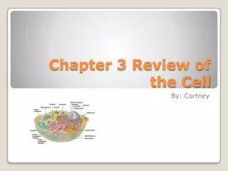
Chapter 3 Review of the Cell. By: Cortney. In 1665, a scientist named Robert Hooke first described what cells really were. He did this by finding thousands of tiny chambers in dried cork under an early light microscope. After the next 175 years he research led to the Cell Theory.

E N D
Chapter 3 Review of the Cell By: Cortney
In 1665, a scientist named Robert Hooke first described what cells really were. He did this by finding thousands of tiny chambers in dried cork under an early light microscope. \After the next 175 years he research led to the Cell Theory. • Cell Theory is said to be the most important components of all plant and animal tissues. There are several concepts to the Cell Theory. • Cells are the building blocks of all plants and animals. • Cells are produced by the division of preexisting cells. • Cells are the smallest units that perform all vital physiological functions. • Each cell maintains homeostasis at the cellular level. • And homeostasis at the tissue, organ, and individual levels reflects the combined and coordinated actions. Basic Concepts of the Cell Theory
Cells float in a watery medium known as the extracellular fluid. • The cell membrane separates the cell contents known as the cytoplasm, from the extracellular fluid. • But the cytoplasm can be further subdivided into a fluid known as the cytosol, and the intercellular structures collectively known as organelles. Fluid Contents with Extracellular Fluid
The cell membrane is the outer boundary of the cell also know as the plasma membrane. • The cell membrane is called the phospholipid bilayer because the phospholipids form two distinct layers. And in each layer the phospholipid molecules lie so that hydrophilic heads are at the surface and the hydrophobic tails are on the inside. • There are four many components of the cell membrane and they are • Phospholipids • Proteins • Glycolipids • Cholesterol The Cell Membrane
The cell membrane is called the phospholipid bilayer because the phospholipids form two distinct layers. And in each layer the phospholipid molecules lie so that hydrophilic heads are at the surface and the hydrophobic tails are on the inside. • General Functions of the cell membrane are: • Physical Isolation • Regulation of exchange with the environment • Sensitivity • Structural support Continued..
Cells in the body work together to maintain homeostasis at the tissue, organ, and system levels. • Communication and coordination activities involve the cell membrane which forms the interface between each cell and its surroundings. • Regulation of the intracellular and extracellular environments are different, and those differences are must be maintained to preserve homeostasis. Cell interactions with the environment
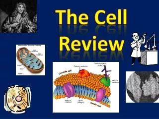
Nonmembranous organelles are always in contact with the cytosol. • These are the Nonmembranous Organelles and their functions: • Cytoskeleton: strength, movement of cellular structures and materials. • Microvilli: Absorption of extracellular materials. • Centrioles: Movement of chromosomes during cell division. • Cilia: Movement of materials over surface. • Ribosomes: Protein synthesis. Nonmembranous Organelles
Membranous organelles are surrounded by lipid membranes that isolate them from the cytosol, just as the cell membrane isolates the cytosol from the extracellular fluid. • These are the Membranous organelles and their functions: • Mitochondria: Produces 95% of the ATP required in a cell. • Nucleus: Control of the metabolism; storage and processing of genetic information. • Nucleolus: Site of the RNA synthesis. • Endoplasmic Reticulum: Synthesis secretory products; intracellular storage and transport. • Rough ER: Secretory protein synthesis. • Smooth ER: Lipid and carbohydrate synthesis. • Golgi Apparatus: Storage, alteration, and packaging secretory products and lysosomes. • Lysosomes: Intracellular removal of damaged or ganelles or of pathogens. • Peroxisomes: Neutralization of toxic compounds. Membranous Organelles
Smooth ER Continued…
The sun is the main component in giving the cell fuel to power all of their functions. • Cells also get energy and power from the mitochondria. • Mitochondria- are small organelles that have an unusual double membrane. • Having both of these energizes that gives the cell al of its energy to fuel all of it functions. Cell Energy
The nucleus is the control center for cellular operations. • The nucleus directs processes that take place in the cytosol and must in turn receive information about conditions and activities in the cytosol. • It also controls the heredity information, control’s the cell’s growth, and reproduction. • Also, it controls the proteins in the cytoplasm and controls the synthesis of ribosomes. Nucleus: The Cell’s Control Center
The G1 phase happens during interphase. The cell is doing its everyday jobs. There are only two chromosomes, and each one has one molecule of DNA. • During S phase the DNA replicates. Now the chromosomes have two molecules of DNA. • In the G2 phase the cell carries out processes that are necessary for mitosis to begin. • Mitosis is the stages where the cell’s DNA gets divided into two separate nuclei. • There are four stages: Prophase, metaphase, anaphase, and telophase. • At the end of the cell’s life cycle, daughter cells are formed. They are each smaller to its parents but are two genetically identical daughter cells. Cell Cycle
The Transmembrane potential is the characteristic of all living cells because it results from the active and passive properties of their cell membranes. • Even though the transmembrane potential is not visible through a microscope, it is just as an important as any other structural characteristic or organelle. • Cell functions that involve the cell membrane involve the transmembrane potential. And because the transmembrane can magnify a stimulus, it greatly increases the cell’s sensitivity to its environment. • The mechanisms that maintain the transmembrane potential is the ions that are rushed into or out of the cell, but it does not depend on the size or nature of the stimulus. Transmembrane Potential
Cells attachment is cells attaching to other cells or to the extracellular protein fibers. These attachments happen at all cell junctions that are not involved in the membrane flow. Also, there are four types of junctions: • Tight Junctions-it blocks the passage of water or solutes between the cells, and is often found where cells are exposed to fluids that are very different from normal extracellular fluids. • Intermediate Junction-is held by a thick layer of proteoglycans that is also called intercellular cement od hyaluronic acid. The cytoplasm at this junction anchor the junction to the cytoskeleton with microfilaments, and help add strength to help stabilize the shape of the cell. • Desmosomes- is very strong in which it can help resist stretching and twisting. It is a superficial layer of skin as well. It also creates dead skin cells that shed in thick sheets rather individually. • Junction Complexes-are the cells that line the digestive tract, respiratory tract, and other passageways. But a single junctional complex of all the other three junctions, but with the tight junction closest to the surface. Cells attached to other cells and body tissues