Download
1 / 30
310 likes | 453 Vues
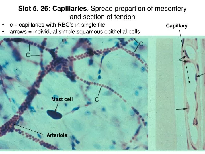
Slot 5. 26: Capillaries . Spread prepartion of mesentery and section of tendon. c = capillaries with RBC’s in single file arrows = individual simple squamous epithelial cells. Capillary. Mast cell. Arteriole. Slot 5.27: Diagram of general structure of blood vessel. Slot 5.28: Artery (l.s.).

E N D
Slot 5. 26: Capillaries. Spread prepartion of mesentery and section of tendon • c = capillaries with RBC’s in single file • arrows = individual simple squamous epithelial cells Capillary Mast cell Arteriole
Slot 5.28: Artery (l.s.) Tunica media (smooth muscle and elastic fibers) Lumen Tunica Intima Inner elastic membrane E=endothelial cells
Slot 5. 29: Arterioles M=tunica media Red blood cells Tunica Intima Internal elastic membrane Endothelial lining Tunica media Tunica adventitia
Slot 5.30: Vein (left) and Artery (right) Tunica intima Tunica media Tunica intima Tunica media Internal elastic membrane Simple squamous Tunica adventitia
L=lymph capillary Arrows = endothelial cells Arrowhead=capillary Artery Muscle tissue Slot 5.31: Lymph capillary
Slot 5.32: Lymph trunk—thoracic duct Tunica intima Tunica media Tunica adventitia Arrows = small blood vessels (vaso vasorum)
Slot 5.34: Lymph node sections P = plasma cells Arrows = large lymphocytes Dashed line = germinal center -Lymph cells dispersed - Lighter staining S= Medullary sinus trabecula Cortex: note crowding of cells Medullary cord
Slot 5.35: Palatine tonsil sections Arrows = arterioles Germinal centers C = mucosal crypt Germinal centers Simple squamous Lymph nodules
Slot 5.36: Thymus T = thymic corpuscles = Hassall’s corpuscles Cortex Medulla Trabeculae divide into lobules
Slot 5.37: Spleen diagram Lymph nodule = white pulp (surrounds central arteriole) Red pulp
Slot 5.38: Arteriole and Venule Tunica media = single layer of smooth muscle Arteriole (lumen circular) Venule
Slot 5.39: Arteriole, Venule, and Lymph capillary Lymph capillaries Venule Larger lymph vessel Arteriole (thicker tunic media)
Tunica adventitia Slot 5.40: Artery and Vein RBC’s Tunica intima Tunica media Vein Artery
Slot 5.41: Artery and Vein Internal elastic membrane Vein Artery
Slot 5.42: Artery Tunica adventitia Internal elastic membrane Artery lumen Endothelial cells Tunica media
Slot 5.43: Artery Tunica intima Tunica media Tunica adventitia Lumen Internal elastic membrane External elastic membrane
Lumen Tunica intima Endothelial lining Tunica media Internal elastic membrane External elastic membrane Tunica adventitia Slot 5.44: Artery
Slot 5.45: Artery Adipose Adipose Vasa vasorum TI Lumen TM TA
Tunica media (w/elastic fibers) Lumen TA Slot 5.46: Aorta
Slot 5.47: Aorta Adipose Tunica media Smooth muscle and fenestrated elastic membranes Tunica adventitia
Slot 5.48: Medium sized vein with elastic fibers Internal elastic membrane Tunica adventitia Tunica media Lumen
Slot 5.49: Lymph Vessel Adipose tissue Valve Flow Lymph vessel
Capsule Slot 5.51: Spleen White pulp Central arterioles Red pulp Trabecula White pulp
Capsule Trabecula Slot 5.52: Spleen Red pulp
Pharynx Slot 5.53: Tonsil (no CT capsule) Stratified squamous epithelial cells Lymph nodule Germinal center (pale)
Slot 5.54: Lymph Node CT capsule Germinal center of lymph nodule Trabecula
Cortex Slot 5.55: Thymus Medulla Hassall’s corpuscles