Download
1 / 16
190 likes | 467 Vues
Testicular Tumours Part 1. Vinod Jain 02.09.2014. Testicular Tumours. Classification Incidence Etiology Spread of tumour Clinical Staging Clinical features Differential Diagnosis Investigations Treatment Follow up schedule. Classification. Primary Tumour. Secondary Tumour.

E N D
Testicular TumoursPart 1 Vinod Jain 02.09.2014
Testicular Tumours Classification Incidence Etiology Spread of tumour Clinical Staging Clinical features Differential Diagnosis Investigations Treatment Follow up schedule
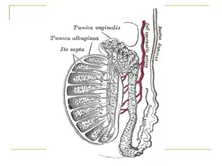
Classification Primary Tumour Secondary Tumour Para testicular neoplasm Lymphona Leukaemia Metastatic Germ Cell tumour Non Germ Celltumour Seminoma (SGCT) Non Semimomatous (NSGCT) Leydig cell Tm Sertoli Cell Tm Gonadoblastoma AdenoCA of rete tests Terratoma Chorio CA Yolk sac Tumour Mixed Tumour EmbryonalCA
Metastatic testicular Tumour In decreasing order Prostate Lung Gut Melanoma Kidney
Incidence • Age – most common solid tumor of men between 20-30 years • Race – White : Black = 4:1 in U.S. • Side – Right > Left • Socio-economic status – high : low = 2:1 • Geographical • Highest in Scandinavia, Germany, Switzerland • Intermediate – USA & UK • Low – Africa and Asia
Age wise incidence of testicular tumour Tumour Type Age group (years) Seminoma 35-40 Pure Terratoma Pediatric age group Embryonal CA 25-30 Chorio CA 25-35 Yolk sac Tumour infancy & child hood Mixed terrato CA 25-30 Lymphoma > 50
Etiology • Congenital – 3-14 times common in undescended testes • Abnormal germ cell morphology • Elevated temperature • Interference with blood supply • Gonadal dysgenesis • Endocrine dysfunction • Acquired • Trauma – co incidence • Endocrine – sex hormone fluctuation • Infection – Mumps induced atrophy/ non-specific infections
Spread of Tumour • Local • Lymphatic – • Right inter aortocaval at L2 precaval preaortic Right common iliac Right ext. iliac • Left Paraortic at renal hilium preaortic common iliac Left ext. iliac (Cross metastasis more common in right side tumour)
Spread of Tumour • Blood (Distant metastases in decreasing order Lung Liver Brain Bone Kidney Adrenal GIT Spleen
Clinical Staging (Boden and Gibbs – 1971) • Stage I (A) – confined to testis with no spread through capsule or spermatic cord • Stage II (B) – Clinical or radiological evidence of spread beyond testis but with in regional L.N. • B1 -<2cm • B2 -2-5cm • B3 - >5cm • Stage III (C) - Disseminated above diaphragm / visceral disease
Clinical features A. Presentations Gradually increasing lump / hardness in testis Abnormal sensitivity – numbness / heaviness / Pain Loss of sexual activity Dull ache in lower abdomen / groin Haemospermia General weakness Metastatic presentations (Contd.)
Clinical features (Contd.) -Metastatic presentations • Cough and Dyspnoea • Anorexia • Nausea / Vomiting (retro duodenal LN) • Neck mass • Swelling lower extremity (IVC obstruction) • Back pain (retroperitoneal L. N.) • Gynaecomastia • Bone pains • Unilateral limb swelling (L.N metastasis) B. Signs • Local • Systemic
Differential Diagnosis Epidedymo-orchitis Testicular haematoma Spermatocele Hydrocele Testicular Torsion
Investigations Haematological – Hb%, Bl. urea/S. creatinine, LFT Tumour markers – AFP, HCG, LDH Scrotal Ultrasound – Usually homogenous, hypoechoic, intra testicular mass X-ray chest CT / MRI – abdomen
Tumour markers NSGCT SGCT AFP N HCG LDH (Advanced) (Advanced)
Let us revise Classification Incidence Etiology Spread of tumour Clinical Staging Clinical features Differential Diagnosis Investigations ---------------------------------------------------------------------------------- Treatment Follow up schedule