Endonuclease cut
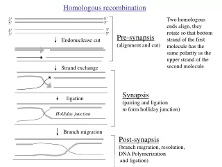
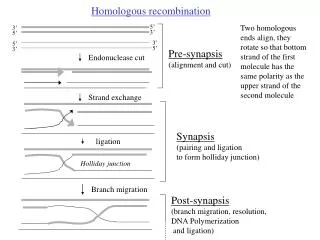
Homologous recombination. 5’. Two homologous ends align, they rotate so that bottom strand of the first molecule has the same polarity as the upper strand of the second molecule. 3’. 3’. 5’. 3’. 5’. 5’. 3’. Pre-synapsis (alignment and cut). Endonuclease cut. Strand exchange. Synapsis

Endonuclease cut
E N D
Presentation Transcript
Homologous recombination 5’ Two homologous ends align, they rotate so that bottom strand of the first molecule has the same polarity as the upper strand of the second molecule 3’ 3’ 5’ 3’ 5’ 5’ 3’ Pre-synapsis (alignment and cut) Endonuclease cut Strand exchange Synapsis (pairing and ligation to form holliday junction) ligation Holliday junction Branch migration Post-synapsis (branch migration, resolution, DNA Polymerization and ligation)
Post-synapsis Resolution of the Holliday junction rotation
Conjugation in E.coli K12 (Transfer of sex plasmid F from F+ cells to F- cells via a pilus) Black= genomic DNA Blue= plasmid F+ X F- Transfer of the newly replicated plasmid into F- bacteria through. Plasmid replicates through rolling circle mechanism F+ F- F+ F+ Occasionally F plasmid integrates into the bacterial genome F+ Hfr
Transfer of newly replicated genome from Hfr to F- X Hfr F- Only partial transfer of F plasmid DNA occurs, as a result F- strain is not converted to Hfr strain Recombinants Homologous recombination
Hfr x F-: a system for isolating genes involved in homologous recombination. Types of mutant screens • Loss of function (Hypo-Rec mutants): • 1. Reduced ability of F minus strain to produce recombinants • when mated with Hfr lawn • 2. Sensitivity to mitomycin C (a DNA damaging drug) • Sensitivity to UV • B. Gain of function (suppressor mutations): • mutations that increase the recombination proficiency of otherwise hypo-rec mutant.
recA (involved in synapsis) RecA protein requires ATP for binding to ssDNA rapidly and cooperatively to generate a nucleo-protein filament (RecA bound ssDNA). The nucleo-protein filament binds non-specifically to dsDNA to generate DNA network. In these networks, ssDNA synapses with homologous dsDNA sequence. Thus, RecA promotes pairing and strand exchange. recB, recC, recD (pre-synapsis) RecB, RecC and RecD proteins are components of RecBCD enzyme, which is also called as ExoV. RecBCD has an ATP dependent dsDNA and ssDNA exonuclease activity. It has an ATP-dependent unwinding activity on linear dsDNA producing ssDNA tails or long ssDNA. During unwinding from right to left makes ss nicks near chi (crossover hotspot instigators) site (5’-GCTGGTGG-3’). It also contains D-loop cleavage activity.
recE (pre-synapsis) Also called as ExoVIII. Has ATP-independent exonuclease activity. Degrades dsDNA from 5’ end, producing long 3’ tails. It normally stays suppressed by the action of sbcA protein. recE gene is activated in recBCD, sbcA mutants. Thus RecE-mediated recombination is a secondary pathway. recF (pre-synapsis) Catalytic activity of RecF is unknown. It probably acts as a switch to divert ssDNA from being replication templates to being a recombination substrates.
Post-synapsis genes for Homologous Recombination RuvAB - These enzymes catalyze branch migration, resulting in pairing of two homologous strands of DNA. RuvAB has DNA helicase activity that causes branch migration from a crossover in the direction that will extend the DNA heteroduplex. RuvC - These enzymes are Holliday junction specific endonucleases that cut two strands of DNA at a "cross-over" (Holliday junction). The resulting DNA strands can be rejoined by DNA ligase, completing the recombination reaction. RuvC interacts with RuvAB to form a RuvABC complex that facilitates the branch migration and recombination reaction.
Pathways of homologous recombination • RecBCD pathway: predominant. • RecFOR pathway: secondary • RecE pathway ? (Differ in pre-synaptic steps. Have mechanistically identical synaptic and post-synaptic steps) RecBCD pathway The RecBCD complex initiates recombination between two homologous dsDNA molecules when one of the molecules has a free, double-stranded end. The RecBCD complex binds to the free end and rapidly degrades the DNA. When RecBCD encounters a short DNA sequence called a chi site, the RecD subunit is altered such that the double-stranded exonuclease is inactivated -- the RecBC complex is converted to a helicase with 5' to 3' single-stranded nuclease activity that unwinds the donor DNA and degrades one strand of the DNA. In addition, the RecB subunit helps "load" RecA protein onto the 3' end of the resulting ssDNA. The ssDNA-RecA complex can invade a homologous dsDNA molecule. (There are other enzymes (RecFOR pathway) in bacteria that can catalyze similar functions, so recBC mutants are not completely defective for recombination).
RecFOR pathway - The RecFOR pathway was discovered by looking for suppressors of recBC mutants (called sbcBCD). RecF binds DNA and has a weak ATPase activity that is stimulated by RecR. RecO promotes the exchange of single-stranded DNA binding protein (SSB) with RecA protein.
RecBCD pathway RecA
Relevance to crop genetic engineering • Gene integration via homologous recombination (HR) between the introduced DNA and the host chromosome will produce the most desirable transgenic product. However, HR-mediated foreign gene integration does not occur efficiently in higher plants. One of the burning topics in plant biology is to develop a strategy for obtaining HR- mediated transgene integration. • Basic features of HR mechanism and the essential proteins (eg. RecA) are conserved between the kingdoms. recA-like genes are found in yeast, animals and plants. recA-like genes of yeast are called Rad51 and Dmc1. Plant homologs of dmc1 have been identified. E. coli genetics Yeast genetics Plant / Animal genetics (conjugation) (mitosis/meiosis) (mitosis/meiosis)
Yeast proteins involved in homologous recombination (Mitosis/ Meiosis) Genetic screen= sensitivity of yeast cells to X-rays RecA counterpart Rad51, Rad52, Rad54, Rad55, and Rad57: These proteins probably complex together to form a recombinosome. Meiosis specific Rad51 is called DMC1. Rad51 is a RecA homolog. Thus, Rad51 in the recombinosome is involved in ATP-dependent strand exchange RecBCD, F, E counterparts Rad50, Mre11, Xrs2: Make a complex that is involved in the structural maintenance of recombination substrate and processing of DNA ends by a 5’—3’ exonulease activity. Rad50 and Mre11 are also implicated to have roles in the illegitimate recombination. Mre11 mutants of chicken show increased homologous recombination frequency (probably due to the suppression of illegitimate recombination)
Strategies for obtaining efficient HR in plants 1. Over-expression of E. coli genes involved in HR: [Bernd Reiss et al. PNAS | March 28, 2000 | vol. 97 | no. 7 | 3358-3363 ] Over-expression of RecA: stimulated sister chromatid exchanges,increased intra-chromosomal recombination but not the frequency of gene targeting (GT is defined as recombination between foreign gene carrying homologous sequences and the complementary genomic sequences). Over-expression of RuvC (RuvCis involved in the resolution of Holliday junction): [Shalev et al. PNAS Vol. 96, Issue 13, 7398-7402 1999] Increased intra-, extra-, and inter-chromosomal recombination but not GT frequency. 2. Modification of plant HR machinery: RAD50 mutant: RAD50 may be a common factor of illegitimate recombination and HR. RAD50 is dispensable for HR. RAD50 plant mutant may have suppressed illegitimate recombination. Intra-chromosomal recombination indeed goes up in RAD50 plant mutant. GT frequency is yet to be demonstrated in RAD50 mutants. 3. Create ds breaks (DSB) in the target site:This is the most successful strategy. It is based on the finding that a ds break (DSB) in plant genome is preferentially repaired by HR process. This led to development of strategies for creating site-specific DSB, such as Zinc-Finger Nuclease-mediated double-stranded break induction.
Zinc-Finger Nuclease (ZFN) Technology Zinc finger nucleases (ZFNs) are a class of engineered DNA-binding proteins that facilitate targeted editing of the genome by creating double-strand breaks in DNA at user-specified locations Invention of ZFN (fundamental discoveries from the lab of S. Chandrasegaran)
Srinivasan Chandrasegaran and Jeff Smith (1999) Biol. Chem., Vol. 380, pp. 841 – 848. Minireview: Chimeric Restriction Enzymes: What Is Next? There are numerous bacterial enzymes that recognize an asymmetric sequence and cleave a short distance from that sequence. These are termed type IIS enzymes, where ‘s’ stands for shifted cleavage. These enzymes do not recognize any specific sequence at the site cut. For example, FokI restriction endonuclease recognizes the non-palindromic penta-deoxyribonucleotide 5’-GGATG-3’:5’-CATCC-3’ in duplex DNA and cleaves 9/13 nucleotides downstream of the recognition site. This property implies the presence of two separate protein domains within FokI: one for sequence-specific recognition of DNA and the other for the endonuclease activity. Once the DNA-binding domain is anchored at the recognition site, a signal is transmitted to the endonuclease domain, probably, through allosteric interactions, and cleavage occurs. We reasoned that the type IIS enzymes probably are the ideal candidates for changing sequence specificities because one may be able to swap the recognition domain of these enzymes with other naturally occurring DNA-binding proteins which recognize longer sequences. Therefore, we undertook a detailed study of the FokI restriction-modification system from Flavobacterium okeanokoites R= recognition or DNA binding domain EN= endonuclease domain
Srinivasan Chandrasegaran and Jeff Smith (1999) Biol. Chem., Vol. 380, pp. 841 – 848. Minireview: Chimeric Restriction Enzymes: What Is Next? The modular nature of FokI endonuclease suggested that it might be feasible to construct chimeric restriction enzymes with novel sequence-specificities by linking other DNA-binding proteins to the cleavage domain of FokI endonuclease. This indeed proved to be the case. We reported the construction of the first ‘chimeric’ restriction endonuclease by linking the Drosophila Ubx homeodomain to the cleavage domain of FokI (Kim and Chandrasegaran, 1994). We then reported the creation of novel site-specific endonucleases by linking two different three zinc-finger proteins to the FokI cleavage domain (Kim et al., 1996). Recently, we reported the creation of a novel site-specific endonuclease by linking the N-terminal 147 amino acids of the yeast Gal4 protein to the cleavage domain of FokI (Kim et al., 1998). Thus, we have shown that the three common eukaryotic DNA-binding motifs, namely the helix-turn-helix motif, the zinc finger motif and the basic helix-loop-helix protein containing a leucine zipper motif, can be converted into novel site-specific endonucleases by fusing them to the FokI cleavage domain. Such engineered chimeric nucleases have been shown to make specific cuts in vitro very close to the expected recognition sequences. Of these chimeric nucleases, the most important are those based on zinc-finger DNA-binding motifs. Because of their modular nature, the zinc finger proteins offer an attractive framework for designing chimeric restriction enzymes with tailor-made sequence-specificities. The Cys2-His2 zinc finger proteins are a class of DNA-binding proteins that contain sequences of the form (Tyr, Phe)-Xaa-Cys-Xaa2-4-Cys-Xaa3-Phe-Xaa5-Leu-Xaa2-His-Xaa3-5-His, usually in tandem arrays. Xaa represents an unspecified amino acid. Each of these sequences binds a Zinc(II) ion to form the structural domain termed a zinc finger. These proteins bind to DNA by inserting an α-helix into the major groove of the double helix.