Download

1 / 41
410 likes | 592 Vues
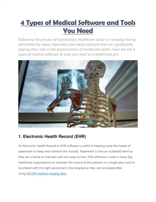
Life Cycle of Medical Imaging Data. Valerie Humblet, PhD Harvard Catalyst Imaging Consortium. What is an image ?. What is an image ?. 2D array of pixels. Pixel dimensions. The pixel size is the dimension in millimeters of the pixels. Life cycle. Clinical need Acquisition Storage

E N D
http://catalyst.harvard.edu Life Cycle of Medical Imaging Data Valerie Humblet, PhD Harvard Catalyst Imaging Consortium
What is an image ? 2D array of pixels
Pixel dimensions The pixel size is the dimension in millimeters of the pixels
Life cycle Clinical need Acquisition Storage Visualization Analysis Registration Segmentation
Life cycle Clinical need Acquisition Storage Visualization Analysis Registration Segmentation
Clinical need Clinical need Diagnosis: Heart disease Stroke Cancer Decision maker: Patient with myocardial infarction, MRI to assess viability for bypass surgery
Life cycle Clinical need Acquisition Storage Visualization Analysis Registration Segmentation
Acquisition Image: Toshiba Image: Siemens Computed Tomography Image: Philips PET/CT Ultrasound Image: BWH Magnetic Resonance Imaging
Data representation Result of acquisition is a 3D volume of data related to the patient The 3D volume is sampled on a 3D grid in the acquisition coordinate system (I,J,K).
Life cycle Clinical need Acquisition Storage Visualization Analysis Registration Segmentation
Storage All medical imaging data is collected and stored in standard radiological file format Digital Imaging and Communications in Medicine DICOM 3.0 (1993) File has complex header that contains fields critically important to image analysis as well as fields with IHI
DICOM 3.0 Data Header 0002,0000,File Meta Elements Group Len=148 0002,0001,File Meta Info Version=256 0002,0002,Media Storage SOP Class UID=1.2.840.10008.5.1.4.1.1.4. 0002,0003,Media Storage SOP Inst UID=0.0.0.0. 0002,0010,Transfer Syntax UID=1.2.840.10008.1.2.1. … 0008,0060,Modality=MR 0008,0070,Manufacturer=GE MEDICAL SYSTEMS 0008,0080,Institution Name=1852796513 0008,0081,City Name=1852796513 0008,0090,Referring Physician's Name=1852796513 0008,0092,?=1852796513 0008,0201,?=-0500 0008,1010,Station Name=1852796513 0008,1030,Study Description=anon 0008,103E,Series Description=anon 0008,1040,Institutional Dept. Name=1852796513 0008,1050,Performing Physician's Name=1852796513 0008,1060,Name Phys(s) Read Study=1852796513 0008,1070,Operator's Name=anon 0008,1080,Admitting Diagnosis Description=1852796513 0008,1090,Manufacturer's Model Name=GENESIS.SIGNA ….. …….. 0028,0010,Rows=256 0028,0011,Columns=256 0028,0030,Pixel Spacing=0.937500 0.937500 0028,0100,Bits Allocated=16 0028,0101,Bits Stored=16 0028,0102,High Bit=15 0028,0103,Pixel Representation=1 ……. 7FE0,0010,Pixel Data=131072
DICOM 3.0 Data Header 0002,0000,File Meta Elements Group Len=148 0002,0001,File Meta Info Version=256 0002,0002,Media Storage SOP Class UID=1.2.840.10008.5.1.4.1.1.4. 0002,0003,Media Storage SOP Inst UID=0.0.0.0. 0002,0010,Transfer Syntax UID=1.2.840.10008.1.2.1. … 0008,0060,Modality=MR 0008,0070,Manufacturer=GE MEDICAL SYSTEMS 0008,0080,Institution Name=1852796513 0008,0081,City Name=1852796513 0008,0090,Referring Physician's Name=1852796513 0008,0092,?=1852796513 0008,0201,?=-0500 0008,1010,Station Name=1852796513 0008,1030,Study Description=anon 0008,103E,Series Description=anon 0008,1040,Institutional Dept. Name=1852796513 0008,1050,Performing Physician's Name=1852796513 0008,1060,Name Phys(s) Read Study=1852796513 0008,1070,Operator's Name=anon 0008,1080,Admitting Diagnosis Description=1852796513 0008,1090,Manufacturer's Model Name=GENESIS.SIGNA ….. 0010,0010,Patient's Name=anon 0010,0020,Patient ID=anon 0010,0030,Patient Date of Birth=00000000 0010,0032,Patient Birth Time=000000 0010,0040,Patient Sex=O 0010,1010,Patient Age=000Y …….. 0028,0010,Rows=256 0028,0011,Columns=256 0028,0030,Pixel Spacing=0.937500 0.937500 0028,0100,Bits Allocated=16 0028,0101,Bits Stored=16 0028,0102,High Bit=15 0028,0103,Pixel Representation=1 Physician and Study information
DICOM 3.0 Data Header 0002,0000,File Meta Elements Group Len=148 0002,0001,File Meta Info Version=256 0002,0002,Media Storage SOP Class UID=1.2.840.10008.5.1.4.1.1.4. 0002,0003,Media Storage SOP Inst UID=0.0.0.0. 0002,0010,Transfer Syntax UID=1.2.840.10008.1.2.1. … 0008,0060,Modality=MR 0008,0070,Manufacturer=GE MEDICAL SYSTEMS 0008,0080,Institution Name=1852796513 0008,0081,City Name=1852796513 0008,0090,Referring Physician's Name=1852796513 0008,0092,?=1852796513 0008,0201,?=-0500 0008,1010,Station Name=1852796513 0008,1030,Study Description=anon 0008,103E,Series Description=anon 0008,1040,Institutional Dept. Name=1852796513 0008,1050,Performing Physician's Name=1852796513 0008,1060,Name Phys(s) Read Study=1852796513 0008,1070,Operator's Name=anon 0008,1080,Admitting Diagnosis Description=1852796513 0008,1090,Manufacturer's Model Name=GENESIS.SIGNA ….. 0010,0010,Patient's Name=anon 0010,0020,Patient ID=anon 0010,0030,Patient Date of Birth=00000000 0010,0032,Patient Birth Time=000000 0010,0040,Patient Sex=O 0010,1010,Patient Age=000Y …….. 0028,0010,Rows=256 0028,0011,Columns=256 0028,0030,Pixel Spacing=0.937500 0.937500 0028,0100,Bits Allocated=16 0028,0101,Bits Stored=16 0028,0102,High Bit=15 0028,0103,Pixel Representation=1 Patient information
DICOM 3.0 Data Header 0002,0000,File Meta Elements Group Len=148 0002,0001,File Meta Info Version=256 0002,0002,Media Storage SOP Class UID=1.2.840.10008.5.1.4.1.1.4. 0002,0003,Media Storage SOP Inst UID=0.0.0.0. 0002,0010,Transfer Syntax UID=1.2.840.10008.1.2.1. … 0008,0060,Modality=MR 0008,0070,Manufacturer=GE MEDICAL SYSTEMS 0008,0080,Institution Name=1852796513 0008,0081,City Name=1852796513 0008,0090,Referring Physician's Name=1852796513 0008,0092,?=1852796513 0008,0201,?=-0500 0008,1010,Station Name=1852796513 0008,1030,Study Description=anon 0008,103E,Series Description=anon 0008,1040,Institutional Dept. Name=1852796513 0008,1050,Performing Physician's Name=1852796513 0008,1060,Name Phys(s) Read Study=1852796513 0008,1070,Operator's Name=anon 0008,1080,Admitting Diagnosis Description=1852796513 0008,1090,Manufacturer's Model Name=GENESIS.SIGNA ….. 0010,0010,Patient's Name=anon 0010,0020,Patient ID=anon 0010,0030,Patient Date of Birth=00000000 0010,0032,Patient Birth Time=000000 0010,0040,Patient Sex=O 0010,1010,Patient Age=000Y …….. 0028,0010,Rows=256 0028,0011,Columns=256 0028,0030,Pixel Spacing=0.937500 0.937500 0028,0100,Bits Allocated=16 0028,0101,Bits Stored=16 0028,0102,High Bit=15 0028,0103,Pixel Representation=1 Image information
DICOM 3.0 Data Header 0002,0003,Media Storage SOP Inst UID=0.0.0.0. 0002,0010,Transfer Syntax UID=1.2.840.10008.1.2.1. … 0008,0060,Modality=MR 0008,0070,Manufacturer=GE MEDICAL SYSTEMS 0008,0080,Institution Name=1852796513 0008,0081,City Name=1852796513 0008,0090,Referring Physician's Name=1852796513 0008,0092,?=1852796513 0008,0201,?=-0500 0008,1010,Station Name=1852796513 0008,1030,Study Description=anon 0008,103E,Series Description=anon 0008,1040,Institutional Dept. Name=1852796513 0008,1050,Performing Physician's Name=1852796513 0008,1060,Name Phys(s) Read Study=1852796513 0008,1070,Operator's Name=anon 0008,1080,Admitting Diagnosis Description=1852796513 0008,1090,Manufacturer's Model Name=GENESIS.SIGNA ….. 0010,0010,Patient's Name=anon 0010,0020,Patient ID=anon 0010,0030,Patient Date of Birth=00000000 0010,0032,Patient Birth Time=000000 0010,0040,Patient Sex=O 0010,1010,Patient Age=000Y …….. 0028,0010,Rows=256 0028,0011,Columns=256 0028,0030,Pixel Spacing=0.937500 0.937500 0028,0100,Bits Allocated=16 0028,0101,Bits Stored=16 0028,0102,High Bit=15 0028,0103,Pixel Representation=1 ……. 7FE0,0010,Pixel Data=131072 The data
Data compression 2 types of algorithms The lossless compression techniques allow the exact original data to be reconstructed from the compressed data. Ex: JPEG-LS The lossy compression techniques deliberately discard information that is not diagnostically important Ex: JPEG
Picture Archiving and Communication System Hospital based computer networked storage of images DICOM images –available immediately Ultrasound, CT, X-ray, MRI, PET, Nuclear medicine Archiving system, replacing hardcopies, CDs, DVDs, accessible through network PACS
Life cycle Clinical need Acquisition Storage Visualization Analysis Registration Segmentation
Visualization Interact in 3D to enhance data interpretation
Anatomical planes The 3D viewer displays a model of the head The 2D viewer display the three anatomical planes (axial, sagittal,coronal)
Axes for spatial coordinates RAS: Right-Anterior-Superior The index i in the file increases from the Left to the Rightside of the patient. The index j in the file increases from Posterior to Anterior. The index k in the file increases from Inferiorto Superior.
Axes for spatial coordinates LPS: Left-Posterior-Superior The index i in the file increases from the Right to the Leftside of the patient. The index j in the file increases from Anterior to Posterior. The index k in the file increases from Inferior to Superior
Life cycle Clinical need Acquisition Storage Visualization Analysis Registration Segmentation
Registration Registration is the process of transforming three different spaces into a common reference frame
Use for image registration Within or Intra-subject: Purpose: To combine functional and anatomical info of different imaging modalities Examples: CT and MR for surgical planning MR and PET/SPECT images of tracer uptake for localization of functional activity Acrossor Inter-subject: Purpose: To assess individual or group variability in some anatomical or functional measure Examples: Responders vs. non-responders to treatment
Use for image registration Serial or Longitudinal: a sequence of images collected over time of the same subject(s) Purpose: To assess change within a subject or group over time due to development, aging, disease progression and/or to monitor response to treatment. Examples: Deformation based morphometry (months), fMRI (minutes) Subject to atlas: Purpose: To use population based information as priors to inform labeling or registration Examples: Brain segmentation and parcellation, Boundary based registration
Why use registration? Same subject, two time points
Compensate for global patient repositioning Preserve distances, planes and angles Appropriate for Bones, brain, optically tracked surgical instrument Often use to initialize non-rigid registration Transformation
Non-rigid transformation Compensate for deformation of the subject
Segmentation Goal: identify or label structures present in the image. Applications: Quantitative measurement of volume, shape or location of structures, Provides boundary for visualization by surface rendering.
Segmentation methods Interactive or manual delineation Supervised approaches with user initialization Thresholding Clustering Region growing Edge detection Atlas based alignment with a template Statistical pattern recognition
Image segmentation Segmentation issues: Interactive segmentation: time consuming. significant intra-rater and inter-rater variability (Warfield et al. 1995). Automatic segmentation: Challenges. Imaging artifacts. Normal and pathological variability. Prospects: Objective assessment of imaging data.
Validation of registration, segmentation or any image processing method Prior to entering clinical practice: Technical validation Speed, robustness, accuracy, reliability Clinical validation Utility, improved diagnosis and patient management FDA approval, incorporation into commercial system Liability
Acknowledgements NIH award #UL1 RR 025758 40