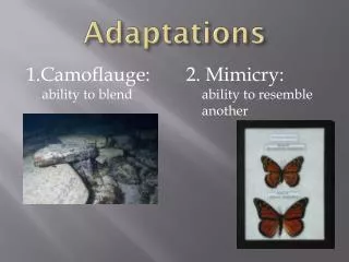
ADAPTATIONS
ADAPTATIONS. PROF. FERDA ÖZKAN MD. To learn how adaptive changes occur To learn the types of adaptive responses. ADAPTATION to INJURY or STRESS. P hysiologic stresses or some pathologic stimul i cause a number of physiologic and morphologic cellular adaptations and abnormal cell growth.


ADAPTATIONS
E N D
Presentation Transcript
ADAPTATIONS PROF. FERDA ÖZKAN MD.
To learn how adaptive changes occur • To learn the types of adaptive responses
ADAPTATION to INJURY or STRESS Physiologic stresses or some pathologic stimuli cause a number of physiologic and morphologic cellular adaptations and abnormal cell growth.
ADAPTATION to INJURY or STRESS Abnormal cellular growth may result in either a decrease or an increase in the mass of the involved tissue.
ATROPHY • Atrophy is a decrease in the size of a tissue or organ, resulting from a decrease either in the size of individual cells or in the number of cells composing the tissue. • Shrinkage in the size of the cell by loss of cell substance is known as atrophy. • Atrophyis distinct from agenesis, aplasia, and hypoplasia, which are abnormalities of organ development.
When a sufficient number of cells are involved, the entire tissue or organ diminishes in size • Atrophy is reversible. • The initial response is a decrease in cell size and organelles, which reduces the metabolic needs of the cell to permit its survival.
In atrophic muscle, the cells contain fewer mitochondria and myofilaments and a reduced amount of rough ER. • Early in the process atrophic cells may have diminished function, but they are not dead. • However, atrophy caused by gradually reduced blood supply may progress to a point at which cells are irreversibly injured and die, usually by apoptosis. • Cell death by apoptosis also contributes to the atrophy of endocrine organs after hormone withdrawal.
MECHANISMS • Atrophy results from decreased protein synthesis and increased protein degradation in cells. • Protein synthesis decreases because of reduced metabolic activity. • The degradation of cellular proteins occurs mainly by the ubiquitin-proteasome pathway. Nutrient deficiency and disuse may activate ubiquitin ligases, which attach the small peptide ubiquitin to cellular proteins and target these proteins for degradation in proteasomes. • This pathway is also thought to be responsible for the accelerated proteolysis seen in a variety of catabolic conditions, including cancer cachexia.
Atrophy is also accompanied by increased autophagy, with resulting increases in the number of autophagic vacuoles. • Autophagy ("self eating") is the process in which the starved cell eats its own components in an attempt to find nutrients and survive. • Autophagic vacuoles are membrane-bound vacuoles that contain fragments of cell components.
Degenerating organelles are taken up in lysosomal vacuoles for enzymatic degradation (autophagy). • Residual organelle membranes (membrane-bound residual bodies) often accumulate in the cytoplasm as brown lipofuscin pigment. • When present in sufficient amounts, they impart a brown discoloration to the tissue (brown atrophy).
ATROPHY • Physiologic atrophy • Pathologic atrophy
ATROPHY • Physiologic atrophy is common during normal development. • Some embryonic structures, such as thyroglossal duct, undergo atrophy during fetal development. • The uterus decreases in size after birth , and this is a form of physiologic atrophy.
The causes of atrophy • 1. Decreased workload (disuse; inactivity) • 2. Loss of innervation (denervation) • 3. Diminished blood supply • 4. Inadequate nutrition • 5. Loss of endocrine stimulation • 6. Pressure • 7. Aging (senility).
Types of atrophy: 1. Regional 2. Systemic
Decreased workload (disuse; inactivity) • Atrophy of disuse occurs in immobilized skeletal muscle and bone, as when a fractured limb is put in a cast or when a patient is restricted to complete bed rest. • Skeletal muscle atrophies rapidly with disuse. • Initially, there is a rapid decrease in cell size that is readily reversible when activity is resumed (simple atrophy). • With more prolonged immobilization, muscle fibers decrease in number ( due to apoptosis) as well as in size (numeric atrophy).
Bone atrophy results when bone resorption occurs more rapidly than bone formation • it is characterized by decreased size of the trabeculae (decreased mass), • Leads to osteoporosis of disuse. • Six-weeks without functional load decreased bone mineral content to 88% of normal.
Loss of innervation (denervation) • Skeletal muscle is dependent on its nerve supply for normal function and structure. • Damage to the lower motor neuron (at any point between the cell body in the spinal cord and the motor end plate) rapid atrophy of the muscle fibers supplied by that nerve • Common in polio, nerve damage, dystrophies. • Physical therapy and electrical stimulation of the muscle
Vascular atrophy (diminished blood supply) A decrease in blood supply (ischemia) to a tissue as a result of slowly developing arterial occlusive disease results in atrophy of the tissue. In late adult life, the brain may undergo progressive atrophy, mainly because of reduced blood supply as a result of atherosclerosis Arterial disease decrease in blood supply (hypoxia) progressive cell loss atrophy • Cerebrovascular disease (atherosclerosis) cerebral atrophy (including neuronal loss).
Inadequate nutrition • Profound protein-calorie malnutrition (marasmus) is associated with the use of skeletal muscle as a source of energy after other reserves such as adipose stores have been depleted. • This results in marked muscle wasting
Loss of endocrine stimulation -endocrine atrophy • The sex glands shrink with depletion of endocrine stimulation. • The endometrium, breast, and many endocrine glands are dependent on trophic hormones. Withdrawal of these hormones leads to atrophy: • When estrogen secretion by the ovary decreases at menopause, there is physiologic atrophy of the endometrium, vaginal epithelium, and breast.
Decreased secretion of pituitary trophic hormones atrophy of the thyroid, adrenals, and gonads. • High-dose adrenal corticosteroid therapy suppression of pituitary corticotropin (adrenocor-ticotrophic hormone-ACTH) secretion atrophy of the adrenal glands. • Withdrawal of steroid therapy in such patients must be gradual enough to permit regeneration of the atrophied adrenal.
Pressure atrophy • Prolonged compression of tissue causes atrophy. • A largeneoplasm in the spinal canal may produce atrophy in both the spinal cord it compresses and the surrounding vertebrae. • Obstruction of the duct of an exocrine gland (sialotilhiasis) atrophy in salivary gland • Obstruction of any ureter atrophy in kidney hydronephrosis.
Genetic atrophy Inherited brain syndromes • cerebral atrophy, • neuronal loss.
Systemic atrophy Atrophy Due to Lack of Nutrients: • Severe protein-calorie malnutrition(marasmus) • marked muscle atrophy • Starvation • disasters • wars • immigrants • GIS pathology (chronic ulceraltive lesions) • poor populations.
Senile Atrophy (Aging) • Cell loss is one of the morphologic changes of the aging process. • It is most apparent in tissues populated by permanent cells, eg, the brain and heart. • Atrophy due to aging is frequently compounded by atrophy due to coexisting factors such as hypoxia.
Atrophy Due to Catabolism&Infections: • Cachexia:catabolism of body tissues, • Infections:AIDS (widespread apoptosis) • Cancer:widespread metastases.
HYPERTROPHY • Hypertrophy results from increased amounts of cytoplasmand cytoplasmic organelles in cells. • In secretory cells, the synthetic apparatus including the endoplasmic reticulum, ribosomes, and the Golgi zone-becomes prominent. • In contractile cells such as muscle fibers, there is an increase in size of cytoplasmic myofibrils.
Increase in the sizes of cells, and hence the size of the organ. Thus, the hypertrophied organ has no new cells, just larger cells. • The increased size of the cells is due not to an increased intake of fluid (cellular swelling). • Hypertrophy may occur as an adaptation to increased demand. • Hypertrophy isreversible.
Physiologic hypertrophy • Hypertrophy of the muscles and heart of a strength athlete • The physiologic growth of the uterus during pregnancy involves both hypertrophy and hyperplasia.
Pathologic hypertrophy • Hypertrophy of the overworked heart of a hypertension patient, or patient of aortic valve stenosis or other cardiac structural defect
Hypertrophy is the result of increased production of cellular proteins • In muscle hypertrophy the linked actions of mechanical sensors (that are triggered by increased work load), growth factors (including TGF-β, insulin-like growth factor-1 [IGF-1], fibroblast growth factor), and vasoactive agents (such as α-adrenergic agonists, endothelin-1, and angiotensin II) are induced. • Mechanical sensors induce production of growth factors and agonists
These stimuli enhance the increase of the synthesis of muscle proteins that are responsible for the hypertrophy. The two main biochemical pathways involved in muscle hypertrophy: • the phosphoinositide 3-kinase/Akt pathway (postulated to be most important in physiologic, e.g., exercise-induced, hypertrophy) and • signaling downstream of G protein-coupled receptors (induced by many growth factors and vasoactive agents, and thought to be more important in pathologic hypertrophy).
HYPERPLASIA • Hyperplasia constitutes an increase in the number of cells in an organ or tissue, which may then have increased volume. • Hypertrophy (increase in cell size) • Hyperplasia (increase in the number of cells). • Hypertrophy does not involve cell division, but hyperplasia takes place if the cellular population is capable of synthesizing DNA, thus permitting mitotic division. • Most hyperplasias seem to result from hormonal stimulation. • As a rule, when the hormone is removed, the hyperplasia quickly reverses.
Physiologic Hyperplasia • (1) hormonal hyperplasia, which increases the functional capacity of a tissue when needed. Hormonal hyperplasia is the proliferation of the glandular epithelium of the female breast at puberty and during pregnancy, usually accompanied by enlargement (hypertrophy) of the glandular epithelial cells. • (2) compensatory hyperplasia increase in tissue mass after damage or partial resection. • The myth of Prometheus, • Punishment for having stolen the secret of fire from the gods, Prometheus was chained to a mountain, and his liver was devoured daily by an eagle, only to regenerate anew every night. • In individuals who donate one lobe of the liver for transplantation, the remaining cells proliferate so that the organ soon grows back to its original size.
Pathologic hyperplasia • Pathologic hyperplasia caused by excesses of hormones or growth factors acting on target cells. Endometrial hyperplasia is an example of abnormal hormone-induced hyperplasia. • In the normal menstruel cycle there is a balance between estrogen and progesterone After a menstrual period proliferative activity in the epithelium is stimulated by pituitary hormones and ovarian estrogen. It is brought to a stop by the rising levels of progesterone, usually about 10 to 14 days before the end of the menstrual period. When the balance between the hormones disturbed with absolute or relative increases in the amount of estrogen end with consequent hyperplasia of the endometrial glands. • This form of pathologic hyperplasia is a common cause of abnormal menstrual bleeding.
Benign prostatic hyperplasia is another common example of pathologic hyperplasia induced by androgens. • Hyperplasia is distinct from cancer, but pathologic hyperplasia constitutesa background for cancerous proliferation
Physiologic Proliferation of the glandular epithelium of the female breast at puberty and during pregnancy Hyperplasia of the pregnant uterus Pathologic The male breast in cirrhosis or estrogen therapy, or in some boys at puberty ("gynecomastia") Adrenal cortex in response to extra ACTH ("stress", ACTH-producing tumor, defective cortisol synthesis) Leydig cells in the testes of men with Klinefelter's syndrome or elderly men Thyroid cells under the influence of hTSH Thyroid cells in response to type V immune injury with stimulation of hTSH receptors ("Grave's disease") Parathyroid cells in renal failure The prostate in old men. Endometrial hyperplasia Hormonal hyperplasia
Physiologic hyperplasia that occurs when a portion of the liver is removed (partial hepatectomy) Prometheus. The callused skin of a laborer's hand. Pathologic Lymph nodes in response to infection Renal tubules in the remaining kidney following removal of the other Bone marrow normoblasts ("erythroblasts") following hemorrhage under the influence of erythropoietin Bone marrow myelocytes in response to infection Bladder epithelial cells in bladder infections. Compensatory hyperplasia
METAPLASIA • Metaplasia is a reversible change in which one mature cell type (epithelial or mesenchymal) is replaced by another mature cell type, and involves thecytoplasm.
Squamous metaplasia • the replacement of airway pseudostratified mucin-producing ciliated columnar epithelium by stratified squamous epithelium (smoking, dust) • transformation of the gallbladder or urinary bladder epithelium to stratified squamous epithelium in the presence of foreign bodies (stones, schistosome eggs).
Columnar mucoid epithelium of theendocervix stratified squamous epithelium in women infected with wart virus (squamous metaplasia), • Replacement of most columnar and transitional epithelium by stratified squamous epithelium (squamous metaplasia), and replacement of corneal epithelium by heavily-keratinized epithelium (vitamin A deficiency),