
ELECTROPHORESIS
E N D
Presentation Transcript
M.Prasad Naidu MSc Medical Biochemistry, Ph.D,. ELECTROPHORESIS
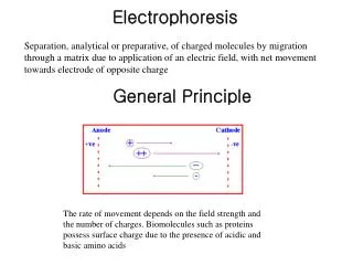
Electrophoresis Electrophoresis may be defined as separation of charged compounds using electric field. Rate of movement of compounds depends upon charge and size of the compound Serum protein electrophoresis – pH8.6 Proteins – negatively charged (an ions) move towards anode
Applications 1. Separation of serum proteins 2. Separation of serum lipoproteins Hyper / Hypo lipoproteinemias 3. Separation of Hemoglobin variants – Hemoglobinopathies
4. Separation iso Enzymes - LDH, CK Classification of Electrophoresis 1. Moving boundary electrophoresis. Described by TISELIUS. He used a U shaped tube and using a buffer separated plasma proteins into 4 fractions, Alb, α, β, γ . Not used now because of poor resolution.
Zone Electrophoresis This is the most commonly used type of electrophoresis. Sample is applied as a band or spot on chemically inert and homogenous support medium on which separation occurs as zones / bands. Based on support medium used zone electrophoresis is sub classified as
1. Paper electrophoresis Cheapest method but the disadvantage is some mixing occurs between different zones. Requires several hours. Separation is not very sharp.
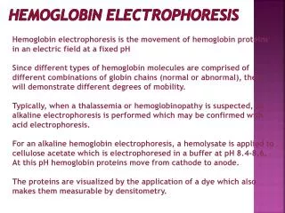
2. Cellulose acetate membrane electrophoresis There is clear separation into discrete zones. Requires about one hour time. 3. Gel electrophoresis A) Agar gel is commonly used for immuno electrophoresis. B) Starch gel is commonly used for separation of iso enzymes
C) Poly acrylamide gel electrophoresis (PAGE). Maximum number of protein bands are obtainted in this technique. Factor affecting separation 1. Charge of the compound 2. Size of the compound
Electrophoresis Unit Electrophoresis chamber consists of two compartments separated from each other by a dividing wall, one side contains the anode and the other the cathode. Each side is filled to the same level with a buffer ( barbital buffer, pH 8.6)
A “bridge” across the top of the dividing wall holds a cellulose membrane or other support material like filter paper, agar gel etc., so that each end of it is in contact with the buffer in one of the compartments. The only connection between the two compartments is by the way of the membrane.
First membrane is immersed in buffer and placed in the chamber and the sample is applied. When a voltage is applied to the cell the current is carried across the porous membrane by the buffer ions.
At. pH 8.6 all the serum proteins carry a net negative charge and tend to migrate toward the anode. Albumin carries the largest charge and therefore moves thefastest. The γ – globulins have the smallest net charge and move the least distance.
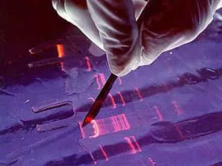
Densitometric Scanning After separation of the compounds by electrophoresis and staining the compounds whether the pattern is normal or abnormal can be visualized. For more accurate fractination densitometry is very valuable
After completion of electrophoresis, the supporting medium is placed in a fixative ( 7 % acetic acid), to prevent diffusion of separated fractions. Separated fractions are then visualized by using appropriate stains, e.g. Bromophenol blue and amido schwartz for plasma proteinsand Sudan black for lipoproteins.
Quantitation is done by Densitometry or Elution, followed by colorimetry or spectro – photometry of the eluted fractions.
- - - - - S
Serum protein electrophoresis Albumin – 55 – 65 % Globulins – 35 – 45%
α1 globulins 2 – 4% α 1 Acid glycoprotein ( oromucoid): 0.6 – 1.4 gm/dl Carbohydrate content is 41%. Oromucoid is considered as an acute phase reactive protein. Increased in acute inflammations & cirrhosis of liver. It binds to progesterone and transports it. Carries carbohydrate to the site of injury for repair.
α1 anti trypsin ( α1 AT) 200 – 400 mg / dl. Active elastase + α1 AT inactive elastase Decreased in emphysema, cirrhosis of liver α 1 Lipoprotein (HDL) AFP ( if present) principal foetal protein normal plasma concentration less than 1 micro gram per dl. Increased in hepatocellular cancer.
α 2 globulins 6 – 12% Haptoglobin - Synthesized in liver. Binds free hemoglobin and prevents its loss through kidney / urine. Heptoglobin concentration is decreased in hemolysis. Caeruloplasmin– contains copper blue coloured protein. Molecular weight 151000. It has 8 sites for copper binding. Normal concentration 30 mg / dl. Decreased in Wilson's disease.
β globulins 8 – 12% Transferrin - Transports iron in plasma. Increased in iron deficiency anemia. Hemo pexin – Binds with heme and prevents its loss through urine β – Lipoprotein (LDL)
γ globulins 12 – 22% Changes in serum protein pattern The Acute Phase Reaction Acute inflammation / tissue necrosis there is increase in serum levels of α1 – acid glycoprotein, α1 AT, ceruloplasmin. Fibrinogen, C – reactive protein and haptoglobin. Increased ESR is due to increased fibrinogen and other globulins.
Para Proteins(M – Proteins) M refers to Myeloma / Monoclonal globulins. para protein may be whole or part of immunoglobulin. If the para protein is of light chain it may spill in to urine (Bence – Jones proteinuria) Bence – jones proteins precipitate around 40 – 600 C and then redissolve at higher temp. These proteins appear as sharp peak ( spike ) mainly in γ region
Causes : Multiple myeloma (Plasma cell myeloma), lymphoma paraprotemias are associated with decreased γ globulins and albumin. Cirrhosis of Liver – Albumin Decreased, γ globulins increased. βγ bridging due to IgA increase
Separation of lipoproteins Chylo - Microns β Preβ LDL α VLDL HDL
Separation of Iso enzymes of LDH MI LDH 1 LDH 2