Bones and Cartilage: Functions, Classification, and Formation
170 likes | 203 Vues
Learn about the functions of bones and cartilage in the human body, their classification by shape, and the process of bone formation. Understand the different types of cartilage and their essential roles in providing support and protection.

Bones and Cartilage: Functions, Classification, and Formation
E N D
Presentation Transcript
Bones and Cartilage prepared by Mr. Abdalmuhaimn Y Sharefcollege of pharmacyHawler medical university20-4-2015
Bones • Functions • Support • Movement: muscles attach by tendons and use bones as levers to move body • Protection • Skull – brain • Vertebrae – spinal cord • Rib cage – thoracic organs • Mineral storage • Calcium and phosphorus • Released as ions into blood as needed • Blood cell formation and energy storage • Bone marrow: red makes blood, yellow stores fat
Classification of bones by shape • Long bones • Short bones • Flat bones • Irregular bones • Sesamoid bones (Short bones include sesmoid bones)
Compact bone : with out cavities. • Spongy bone : with numerous cavities. 2 1
Epiphyses : spongy bone covered by a thin layer of compact bone. Diaphysis : compact bone (totally) with a small component of spongy bone .
Primary bone tissue :Immature : is the first bone tissue to appear in embryonic development . (random disposition of fine collagen fibers) .replaced in adults by? • Secondary bone tissue : collagen fibers arranged in lamellae.
Osteocytes : derive from osteoblasts ,lie in the lacunae between the lamella of matrix.
Isolated osteon: • Nutrients diffuse from vessels in central canal • Alternating direction of collagen fibers increases resistance to twisting forces
outer Volkmans canal do not have concentric lamellae. Osteon communicate with the marrow cavity, the periosteum, and Volkmans canal.
Hematopoiesis Trabeculae have lamellae and osteocytes like osteons
Ossification: bone can be formed in two ways: • Intramembranous ossification : forms directly from mesenchyme (not modeled first in cartilage).direct mineralization of matrix secreted by osteoblasts.(flat bones). • Endochondral ossification: modeled in hyaline cartilage then replaced by bone tissue . All the rest of the bones. • Responsible for the formation of short and long bones.
Intramembranous bone formation • Connective tissue develops embryonically and gives rise to bone • Cells in connective tissue differentiate into osteoblasts • Osteoblasts lay down collagen eventually starts to calcify and cells become osteocytes
Spongy bone • Layers of lamellae and osteocytes • Seem to align along stress lines
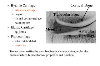
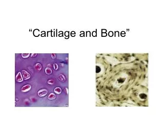
Types of cartilage: 3 • Hyaline cartilage: flexible and resilient • Chondrocytes appear spherical • Lacuna – cavity in matrix holding chondrocyte • Collagen the only fiber • Elastic cartilage: highly bendable • Matrix with elastic as well as collagen fibers • Epiglottis, larynx and outer ear • Fibrocartilage: resists compression and tension • Rows of thick collagen fibers alternating with rows of chondrocytes (in matrix) • Knee menisci and annunulus fibrosis of intervertebral discs
Functions: cartilage • Rigid, yet more flexible than bone; more elastic than bone • Support and protection • Abundant in the fetus and embryo • Site of skeletal growth • Covers joints • Supports nose, ears, trachea, ribs, etc