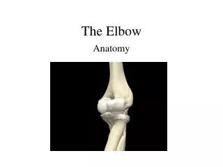
the elbow
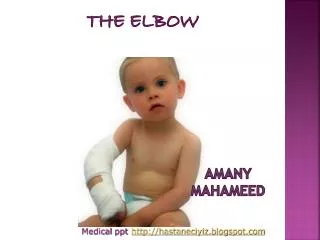
the elbow. AMANy Mahameed. http://hastaneciyiz. blogspot .com. Medical ppt. The important structures of the elbow can be divided into several categories. These include:. Bones and joints Ligaments and tendons Capsule Muscles Nerves Blood vessels. Bones and Joints:.

the elbow
E N D
Presentation Transcript
the elbow AMANyMahameed http://hastaneciyiz.blogspot.com Medicalppt
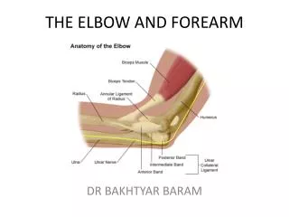
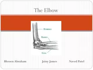
The important structures of the elbow can be divided into several categories. These include: • Bones and joints • Ligaments and tendons • Capsule • Muscles • Nerves • Blood vessels
Bones and Joints: • There are 3 joints at the elbow: 1- Humeroulnar joint (hinge joint) – between trochlea of humerus and trochlea notch of ulnar 2- Humeroradial joint (hinge joint) – between capitilum of humerus and radius head 3- Proximal radioulnar joint (pivot joint)- between proximal end of ulna and proximal end of radius.
Articular cartilage is the material that covers theends of the bones of any joint.with functions to absorb shock and make motion at the elbow joint easier.
Ligaments and Tendons: Ligaments aresoft tissue structures that connect bones to bones. The ligaments around a joint usually combine together to form a joint capsule. • Lateral Collateral Ligament • Medial Collateral Ligament (Together these two ligaments connect the humerus to the ulna, to form the main source of stability for the elbow) • Annular Ligament: that wraps around the radial head and holds it tight against the ulna.
Tendons: • Biceps tendon anteriorly (allows elbow to bend with force) • Triceps tendon posteriorly (allows elbow to straighten with force)
Capsule: • The articular surfaces are connected together by a capsule • Anterior part – from radial and coronoid fossa of humerus to coronoid process of ulna and annular ligament of radius • Posterior part – from capitulum, olecranon fossa, and lateral epicondyle of humerus to annular ligament of radius, olecranon of ulna, and posterior to radial notch.
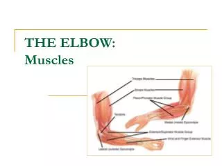
Muslces: • The wrist extensors originate from the lateral epicondyle of the humerus • The wrist flexors originate from the medial epicondyle of the humerus
Nerves: • The median and ulnar nerve are at risk of injury in: • Elbow dislocation • Supracondylar fracture of humerus
Blood vessels: ThEbRartery splits into two branches just below the elbow: the ulnar artery andthe radial artery
Carrying Angle: • The normal carrying angle of the elbow is about 15 degrees of valgus in males and up to 20 degrees in females. It is the angle at which the humerus and forearmarticulate, with the elbow in full extension, and the palms facing forward The carrying angle permits the arm to be swung without contacting the hips www.icareunit.com
Cubitus Varus: “ bow-elbow” Causes Supracondylar fractures of the childhood. *Function of the elbow is almost normal. www.icareunit.com
Treatment: Usually indicated for cosmetic reasons; Consists of removing a bone wedge from the lateral aspect of supracondylar area. www.icareunit.com
Cubitus Valgus: Causes: 1- Errors in management of lateral humeral condyle fracture. When present at birth it can be a sign of Turner or Noonan syndrome. www.icareunit.com
Cubitus Valgus Clinical features • Obvious lateral angulation of the elbow. • Tardy Ulnar Nerve Palsy. (most important sequel( • The ulnar nerve is repeatedly stretched behind the medial epicondyle (for many years) insidious impairment of the nerve trunk with ulnar nerve palsy. www.icareunit.com
Cubitus Valgus Treatment: Elbow deformity needs no treatment, but the nerve palsy is treated by transposing the nerve anterior to the elbow. www.icareunit.com
Tennis Elbow (Lateral Epicondylitis) : • The most common pathology in the elbow. • Lateral epicondylitis (tennis elbow) is an overuse injury involving the extensor muscles that originate on the lateral epicondylar region of the distal humerus (any activity involving extension and/or supination). It is more properly termed a tendinosis that specifically involves the origin of the extensor carpi radialis brevis muscle.
Occurs in up to 50% of tennis players • Cause has been attributed to microscopic tearing with formation of reparative tissue (ie, angiofibroblastic hyperplasia) in the origin of the extensor carpi radialis brevis (ECRB) muscle • Patients present with lateral elbow and forearm pain exacerbated by use • Typical patient- man or woman between 35-55 years who is a recreational athlete or who engages in rigorous daily activities
Treatment: - Rest • Counterforce brace • NSAIDs • Wrist splinting • Corticosteroid and injections • Low level laser therapy
Approximately 90-95% of patients respond to conservative measures and do not require surgical intervention. Patients whose condition is unresponsive to 6 months of conservative therapy (including corticosteroid injections) are candidates for surgery.
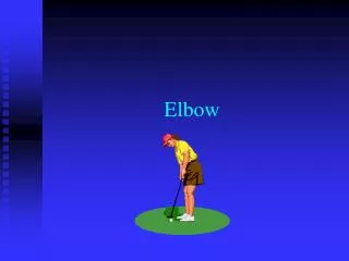
Golfer’s Elbow (Medial Epicondylitis): • This condition is an overuse syndrome that is characterized by pain at the flexor-pronator tendinous origin and is seen in sports activities with repetitive valgus stress, flexion, and pronation, such as occurs in golf, baseball, tennis, fencing, and swimming.This condition is also seen with occupations that require hand, wrist, and forearm motions.
Most common cause of medial elbow pain but less common than tennis elbow • Males: females = 2:1 • Presence of microtears in the flexor-pronator tendons without inflammation • Patient presents with achy pain over the anterior medial epicondyle, usually during activity, and the patient may describe weakness in the forearm or hand. In addition, radiation of the pain may occur in the shoulder, forearm, or hand.
Treatment: 1- Patient education and golf-swing (or the relevant activity) modification 2- Nonsteroidal anti-inflammatory drugs (NSAIDs) 3- Counterforce brace 4- Wrist splints 5 -Corticosteroid injections Surgical treatment should be considered in cases in which conservative treatment has failed after 6-12 months and after all other pathology has been excluded. http://hastaneciyiz.blogspot.com Medicalppt