Download

1 / 24
240 likes | 430 Vues
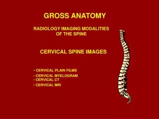
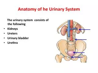
Gross Anatomy of Urinary system. Medical ppt. http://hastaneciyiz.blogspot.com. Urinary system. Functions of Urinary System. Kidneys carry out four functions Filter nitrogenous wastes, toxins, ions, etc. from blood to be excreted as urine.

E N D
Gross Anatomy of Urinary system Medical ppt http://hastaneciyiz.blogspot.com
Functions of Urinary System • Kidneys carry out four functions • Filter nitrogenous wastes, toxins, ions, etc. from blood to be excreted as urine. • Regulate volume and chemical composition of blood (water, salts, acids, bases). • Produce regulatory enzymes. • Renin – regulates BP/ kidney function • Erythropoietin – stimulates RBC production from marrow. • Metabolism of Vitamin D to active form.
Urinary System • Two Kidneys • Perform all functions except actual excretion. • Two Ureters • Convey urine from Kidneys to Urinary Bladder • Urinary Bladder • Holds Urine until excretion • Urethra • Conveys urine from bladder to outside of body
Renal Connective Tissue • Renal fascia- anchors the kidneys to nearby structures. It is dense connective tissue • Adipose capsule/Pararenal Fat Mass of fat tissue that surrounds renal capsule Functions • Keeps kidney in place • Provide cushion effects • Renal capsule- Layer of dense connective tissue that surrounds kidney and supports the soft internal tissues.
Position of the Kidneys • Kidneys are located on either side of the vertebral column: • left kidney lies superior to right kidney • superior surface capped by adrenal gland
Location and Position of the Kidneys • The kidney is positioned between the 12th thoracic and 3rd lumbar vertebrae. • It is retroperitoneal (lies on the posterior abdominal wall, posterior to the peritoneum). • Right kidney is lower than left kidney due to the shape of the liver. • Lateral surface of kidney is convex while medial is concave.
External Anatomy of Kidney • Average size – 12cm x 6cm x 3 cm • Weights 150 grams or 5 oz • Surrounded by three membranes (deep to superficial) • Renal capsule – fibrous barrier for kidneys. • Adipose capsule – fatty tissue designed for protection / stability. • Renal fascia – dense fibrous connective tissue that anchors kidneys/ adrenals to surroundings.
External Anatomy of the Kidney • Lateral surface- convex • Medial surface is concave • Renal Hilum • Indentation where blood vessels, nerves and ureters enter and exit the kidneys. • Renal Sinus • Internal cavity within kidney.
Internal Anatomy of the Kidney • Cortex - Superficial region of kidney. • It is light and granular • Medulla - Deep central region of the kidney. • Deep layer • It is darker • Renal pyramids- Cone shaped structure within the renal medulla. • Base lies against cortex • Apex is the papilla • Renal column- Extensions of cortex that separate renal pyramids within the medulla.
Kidney Internal Anatomy • Renal Pelvis • Flat funnel-shaped expansion of ureter • Major Calices • Large cup-shaped branches of renal pelvis • Minor Calices • Cup-shaped divisions of major calices • Surround papilla of pyramid
Renal Lobe • Consists of: • One renal pyramid • overlying area of renal cortex • adjacent tissues of renal columns
Blood Flow – Arteries • Renal arteries • Segmental arteries • Lobar arteries • Interlobar arteries • Arcuate arteries • Interlobular arteries • Afferent arterioles
Blood Flow – Veins • From nephron • Interlobular veins • Arcuate veins • Interlobar veins • Renal vein
Ureters • Continuation of renal pelvis • Slender tubes that transport urine from kidneys to bladder • Retroperitoneal/Runs behind the peritoneum
Urinary Bladder • Prostate gland: Found in In males • - Lies directly inferior to the bladder • - Surrounds the urethra • A collapsible muscular sac • Stores and expels urine - Full bladder – spherical • Expands into the abdominal cavity - Empty bladder – lies entirely within the pelvis
Urinary Bladder • Muscular sac that stores and expels urine • Location • Pelvic floor • Posterior to public symphysis • Anterior to • Rectum in males • Vagina & uterus in females
Urinary Bladder and Urethra in Male and Female • Trigone of the urinary bladder has three openings; • - Two openings from the ureters • One opening to the urethra
Structure of the Urinary Bladder • Male: • Penis • Dual function • Female: • External urethral orifice
Urethra • In females • Length of 3–4 cm • In males – 20 cm in length – three named regions • Prostatic urethra • Passes through the prostate gland • Membranous urethra • Through the urogenital diaphragm • Spongy (penile) urethra • Passes through the length of the penis
Urethral Sphincter • Internal urethral sphincter • Involuntary smooth muscle • External urethral sphincter • Voluntarily inhibits urination • Relaxes when one urinates
Medical ppt http://hastaneciyiz.blogspot.com