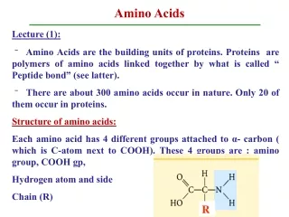
Download
1 / 16
160 likes | 279 Vues
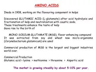
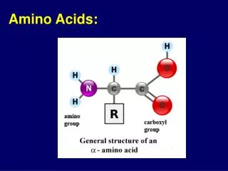
Sources of Amino Acids. De Novo Synthesis CO 2 fixation (ala, asp, glu) little incorporated into protein Host Plasma uptake of all amino acids in vitro growth requires ile, met, cys, gln, glu Digestion of Host Hemoglobin. Hemoglobin. 95% of total erythrocyte protein

E N D
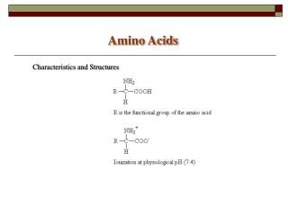
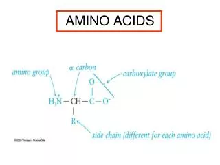
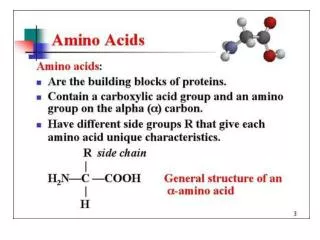
Sources of Amino Acids • De Novo Synthesis • CO2 fixation (ala, asp, glu) • little incorporated into protein • Host Plasma • uptake of all amino acids • in vitro growth requires ile, met, cys, gln, glu • Digestion of Host Hemoglobin
Hemoglobin • 95% of total erythrocyte protein • very abundant (>300 mg/ml or approximately 5 mM) • 60-80% is degraded during erythrocytic stage • 110 g (of 750 total) is consumed in 48 hrs at 20% parasitemia
Endocytosis of Host Cytoplasm cytostome food vacuole pinocytosis (rings)
The Food VacuoleA Specialized Lysosome ATP hemoglobin digestion H+ (pH 5-5.4) ADP • Food Vacuole Proteases • plasmepsins I & II (acid) • falcipains I - III (thiol) • falcilysin (metallo) • Absent: • other acid hydrolases Endocytic Pathway parasite cytoplasm
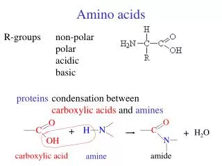
Proteases Mediate the Catabolism of Proteins • proteases (aka peptidases) break the peptide bonds that hold amino acids together • exopeptidases remove amino acids sequentially from either N- or C-terminus • endopeptidases cleave between ‘specific’ residues within polypeptide chain
Initial plasmepsin cleavage is specificand leads to a destabilization of hemoglobin • native Hb is cleaved between Phe-33 and Leu-34 ( chains) • ‘hinge region’ • conserved • important for tetramer stability • the large globin fragments dissociate • heme is released • globin fragments are susceptible to further proteolysis a-F33/L34 í
hemoglobin plasmepsin large globin fragments heme + falcipain plasmepsin medium fragments (20 amino acids) small fragments (6-8 amino acids) falcilysin Hemoglobin Digestion is an Ordered Process • exopeptidase? • free amino acids?
ABC Transporter Super Family • large and ubiquitous gene family • defined by ATP-Binding Cassette • aka Multi-Drug Resistance (MDR) • transport is usually specific for particular types of substrates • Pfmdr-1 protein localized to food vacuole • Pfmdr-1 complements yeast ste6 gene • ste6 transports a-type mating factor (12 residue peptide)
The Food VacuoleA Specialized Lysosome ATP hemoglobin H+ plasmepsin globin fragments ADP heme + amino acids falcipain plasmepsin falcilysin • ABC transporter associated with food vacuole • amino-peptidase activities in parasite cytoplasm amino- peptidase ATP Pfmdr-1? small fragments (6-8 amino acids) ADP
Free Heme is Toxic • heme destabilizes and lyses membranes • hydrolases released into parasite cytoplasm • parasite dies • Possible Detoxification Mechanisms • heme hemazoin (malaria pigment) • H2O2 mediated degradation • GSH mediated degradation • heme oxygenase (P.b. and P.k. only)
Hemazoin = b-Hematin b-hematin heme
b-hematin forms insoluble crystals 'biocrystallization' or 'biomineralization'
Pigment Formation • biocrystallization mechanism unknown • beta-hematin can form spontaneously (harsh conditions) • histidine-rich proteins or lipids can promote the process • heme biocrystallization inhibited by chloroquine and other anti-malarials
The Food VacuoleA Specialized Lysosome ATP hemoglobin H+ plasmepsin Fe2+ O2 ADP globin fragments Fe3+ heme + amino acids -O2 O2 falcipain plasmepsin falcilysin ? • iron oxidized after release from Hb • oxidation promotes formation of ROI • oxidative stress amino- peptidase hemazoin ATP small fragments (6-8 amino acids) Pfmdr-1? ADP
The Food VacuoleA Specialized Lysosome ATP hemoglobin H+ plasmepsin Fe2+ O2 ADP globin fragments Fe3+ heme + amino acids -O2 O2 falcipain plasmepsin falcilysin ? superoxide dismutase? amino- peptidase H2O2 hemazoin ATP catalase? small fragments (6-8 amino acids) Pfmdr-1? H2O + O2 ADP